. 2011;6(12):e28531.
doi: 10.1371/journal.pone.0028531. Epub 2011 Dec 14.

Endoplasmic reticulum stress pathway-mediated apoptosis in macrophages contributes to the survival of Mycobacterium tuberculosis

Affiliations

Endoplasmic reticulum stress pathway-mediated apoptosis in macrophages contributes to the survival of Mycobacterium tuberculosis

Yun-Ji Lim et al. PLoS One. 2011.

Abstract

Background: Apoptosis is thought to play a role in host defenses against intracellular pathogens, including Mycobacterium tuberculosis (Mtb), by preventing the release of intracellular components and the spread of mycobacterial infection. This study aims to investigate the role of endoplasmic reticulum (ER) stress mediated apoptosis in mycobacteria infected macrophages.

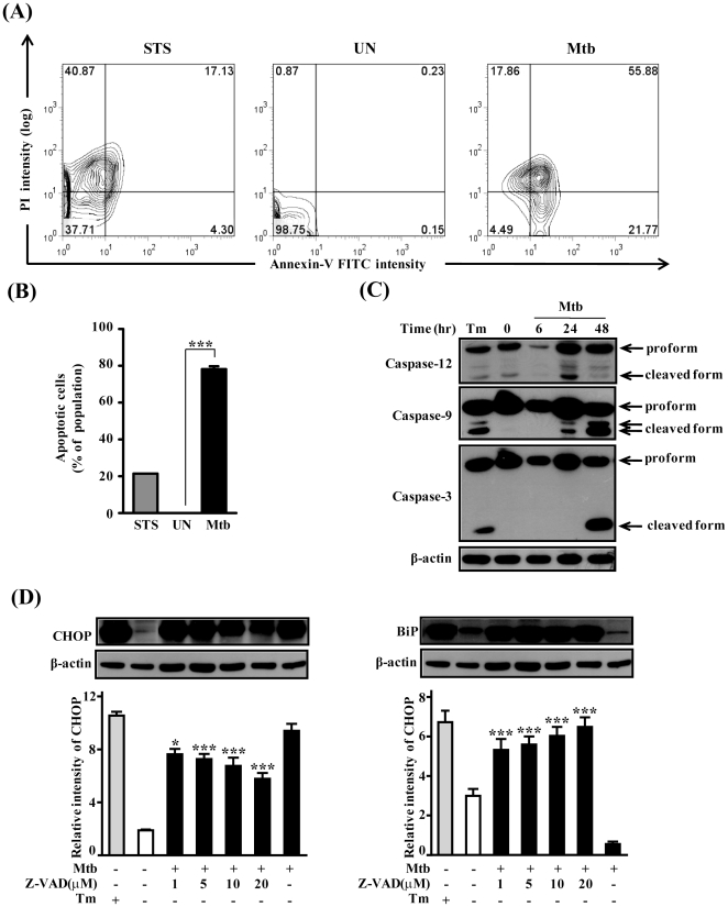
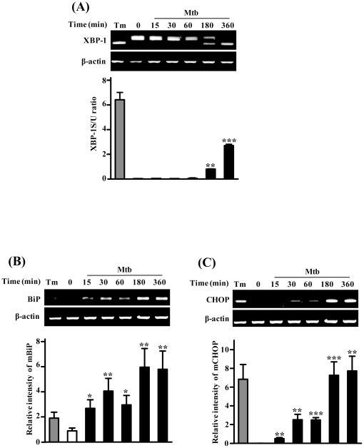
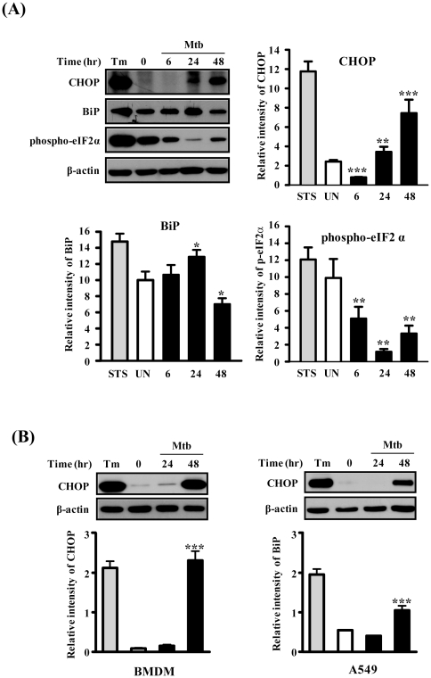
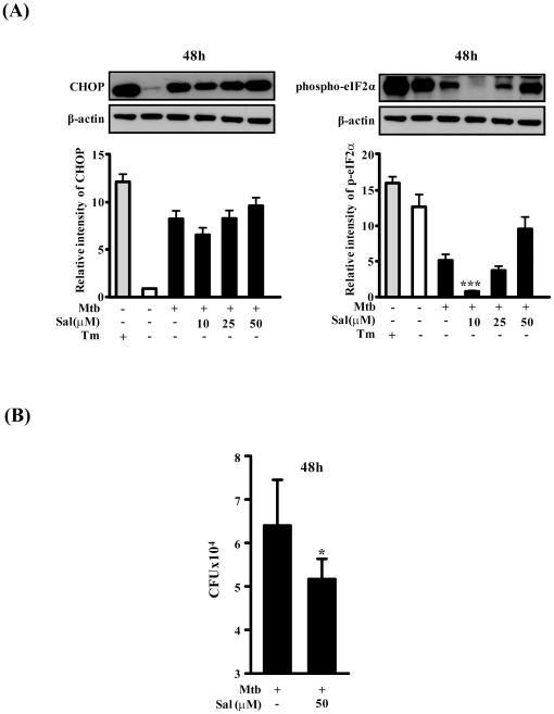
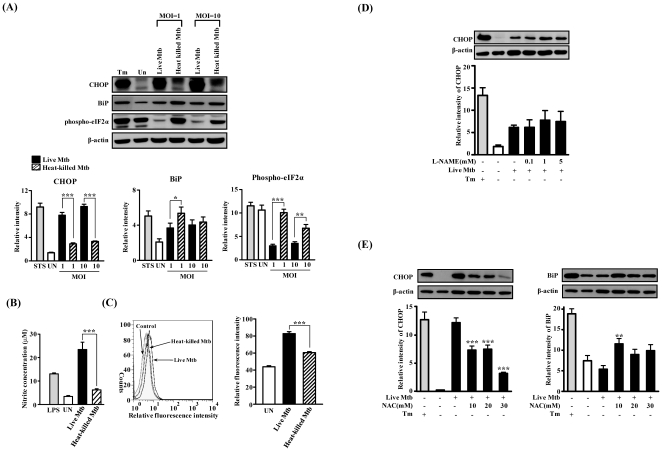
Methodology/principal findings: Here, we demonstrate that ER stress-induced apoptosis is associated with Mtb H37Rv-induced cell death of Raw264.7 murine macrophages. We have shown that Mtb H37Rv induced apoptosis are involved in activation of caspase-12, which resides on the cytoplasmic district of the ER. Mtb infection increase levels of other ER stress indicators in a time-dependent manner. Phosphorylation of eIF2α was decreased gradually after Mtb H37Rv infection signifying that Mtb H37Rv infection may affect eIF2α phosphorylation in an attempt to survive within macrophages. Interestingly, the survival of mycobacteria in macrophages was enhanced by silencing CHOP expression. In contrast, survival rate of mycobacteria was reduced by phosphorylation of the eIF2α. Futhermore, the levels of ROS, NO or CHOP expression were significantly increased by live Mtb H37Rv compared to heat-killed Mtb H37Rv indicating that live Mtb H37Rv could induce ER stress response.

Conclusion/significance: These findings indicate that eIF2α/CHOP pathway may influence intracellular survival of Mtb H37Rv in macrophages and only live Mtb H37Rv can induce ER stress response. The data support the ER stress pathway plays an important role in the pathogenesis and persistence of mycobacteria.


Conflict of interest statement

Competing Interests: The authors have declared that no competing interests exist.

Figures

Figure 1
Figure 1. Mtb H37Rv infection induces apoptosis and caspase activation.
(A) Raw264.7 cells were screened for induction of apoptosis using Annexin-V/PI staining after 48 h infection with Mtb H37Rv at a MOI of 1. Staurosporine (500 nM) was used for making positive control for apoptosis. After washing and Annexin V-/PI staining, cells were analyzed by flow cytometry. Data are respresentative of at least three independent experiments with similar results. (B) Quantitative analysis of the percentage of Annexin V-positive cells as described in B. ***, P<0.001 (C) Cellular levels of caspase-3, caspase-9, and caspase-12 during Mtb-infection in Raw264.7 cells. (D) The effect of caspase inhibitor z-VAD-fmk on the CHOP expression in Mtb-infected Raw264.7 cells. Raw264.7 cells were infected with Mtb at a MOI of 1 for 3 h, and then incubated for 0–48 h. The statistical significance (***P<0.001) of observed differences between z-VAD-fmk treated and untreated groups following infection with Mtb H37Rv were verified by two-tailed t-test. Immunoblot analysis was performed as described in Materials and Methods.
Figure 2
Figure 2. Time-course analysis mRNA for ER stress-related genes after Mtb H37Rv infection.
Raw264.7 cells were infected with Mtb H37Rv (MOI = 1) for 3 h, and then incubated for 0–6 h. (A) XBP-1 mRNA splicing was determined by RT-PCR using specific primers that was used to amplify products of unspliced and spliced mRNA. The results represent the ratio of XBP-1 splicing to XBP-1 unsplicing (XBP-1S/U ratio). (B, C) PCR amplification with the primer pair corresponding to the BiP and CHOP mRNA was performed at the indicated time points. The results were quantified by densitometry. For a positive control, cells were treated with 2.5 µg/mL tunicamycin (Tm) for 6 h. The statistical significance (*P<0.05, **P<0.01 and ***P<0.001) of observed differences between Mtb H37Rv infected and uninfected groups were verified by two-tailed t-test. Representative data from three independent experiments are shown.
Figure 3
Figure 3. Mtb H37Rv infection induces ER stress sensor proteins in macrophage cells and CHOP is activated in BMDMs or A549 cells.
Cells were infected with Mtb H37Rv (MOI = 1) for 3 h, and then incubated for 0–48 h. Immunoblot analysis was performed as described in Materials and Methods. The statistical significance (*P<0.05, **P<0.01 and ***P<0.001) of observed differences between Mtb H37Rv infected and uninfected groups were verified by two-tailed t-test. (A) Raw264.7 cells, (B) BMDMs or A549 cells. Representative data from three independent experiments are shown. STS: staurosporine.
Figure 4
Figure 4. Effect of eIF2α phosphorylation on intracellular survival of Mtb H37Rv.
Raw264.7 cells were pretreated for 30 min with indicated concentrations of salubrinal and then infected with Mtb H37Rv (MOI = 1) for 3 h. Salubrinal remained for the rest of the infection. (A) The cells were incubated for 48 h and Western blot analysis was performed using antibodies directed against CHOP, p-eIF2α and β-actin. DMSO alone was used as the negative control. (B) Quantification of intracellular survival of Mtb H37Rv in Raw264.7 cells pretreated for 30 min with salubrinal as described above. The cells were collected at 48 h postinfection with Mtb H37Rv and bacteria number was determined by CFU counting. The statistical significance (*P<0.05) of observed differences between salubrinal treated and untreated groups following infection with Mtb H37Rv were verified by two-tailed t-test. Data represent the mean±standard error of the mean (SEM) of values obtained in three independent experiments.
Figure 5
Figure 5. Effect of siCHOP on intracellular survival of Mtb H37Rv.
(A) Representative images of Raw264.7 cells after transfection. Raw264.7 cells were transfected with siRNA (siControl, or siCHOP) for 5 h and then infected with Mtb H37Rv for 3 h. At 48 h after Mtb H37Rv infection, Western blot analysis was performed using antibodies directed against CHOP, BiP and β-actin. The experiments were repeated three times. (B) Quantification of intracellular survival of Mtb H37Rv in Raw264.7 cells pretreated with siRNA as described above. The cells were collected at 48 h postinfection with Mtb H37Rv and bacteria number was determined by CFU counting. The statistical significance (**P<0.01) of observed differences between siCHOP treated and untreated groups following infection with Mtb H37Rv were verified by two-tailed t-test. Data represent the mean ± SEM of values obtained in two independent experiments performed in triplicate.
Figure 6
Figure 6. The expression of ER stress markers in Raw264.7 cells infected with live or heat-killed Mtb H37Rv.
Raw264.7 cells were infected with live or heat killed Mtb H37Rv (MOI = 1 to 10) for 3 h, and then incubated for 48 h in the presence or absence of L-NAME or NAC. (A, D, E) Immunoblot analysis was performed as described in Materials and Methods. Representative data from three independent experiments are shown. (B) Effect of Mtb H37Rv infection on nitric oxide (NO) production was assessed indirectly by Griess reaction. (C) Representative flow cytometry histograms of superoxide at 48 h after Mtb H37Rv infection. Superoxide detection was evaluated by dihydroethidium (DHE) staining using flow cytometry. Data are means ± SEM of two independent experiments performed in triplicate. CHOP expression analysis after treatment with N-nitro-L-arginine methyl ester (L-NAME), a nitric oxide inhibitor (D) and N-acetyl-L-cysteine (NAC), a superoxide inhibitor (E). The statistical significance (*P<0.05, **P<0.01 and ***P<0.001) of observed differences between inhibitor treated and untreated groups following infection with Mtb H37Rv were verified by two-tailed t-test.

References

    1. Chiang CY, Centis R, Migliori GB. Drug-resistant tuberculosis: past, present, future. Respirology. 2010;15:413–432. - PubMed
    1. Giacomini E, Iona E, Ferroni L, Miettinen M, Fattorini L, et al. Infection of human macrophages and dendritic cells with Mycobacterium tuberculosis induces a differential cytokine gene expression that modulates T cell response. J Immunol. 2001;166:7033–7041. - PubMed
    1. Pieters J. Evasion of host cell defense mechanisms by pathogenic bacteria. Curr Opin Immunol. 2001;13:37–44. - PubMed
    1. Rachman H, Kim N, Ulrichs T, Baumann S, Pradl L, et al. Critical role of methylglyoxal and AGE in mycobacteria-induced macrophage apoptosis and activation. PLoS One. 2006;1:e29. - PMC - PubMed
    1. Briken V, Miller JL. Living on the edge: inhibition of host cell apoptosis by Mycobacterium tuberculosis. Future Microbiol. 2008;3:415–422. - PMC - PubMed

Publication types

MeSH terms

LinkOut - more resources