. 2011;6(12):e28611.
doi: 10.1371/journal.pone.0028611. Epub 2011 Dec 14.

PEGylation potentiates the effectiveness of an antagonistic peptide that targets the EphB4 receptor with nanomolar affinity

Affiliations

PEGylation potentiates the effectiveness of an antagonistic peptide that targets the EphB4 receptor with nanomolar affinity

Roberta Noberini et al. PLoS One. 2011.

Abstract

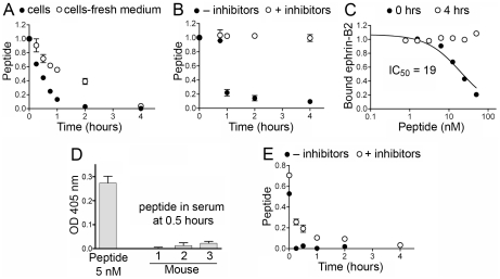
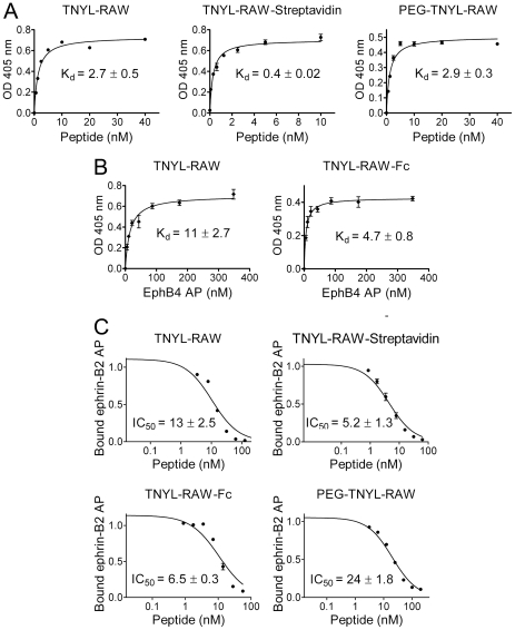
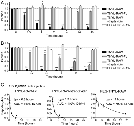
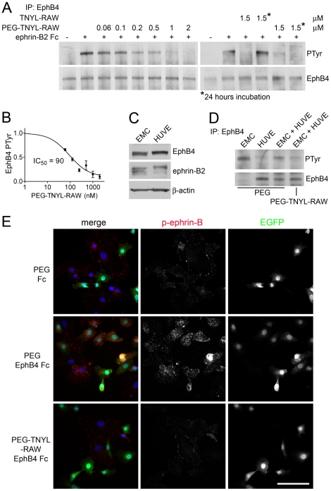
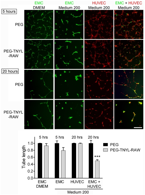
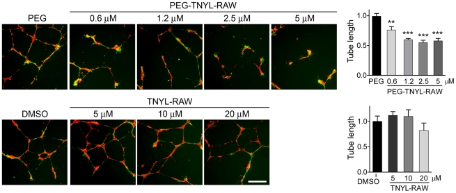
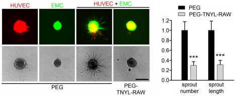
The EphB4 receptor tyrosine kinase together with its preferred ligand, ephrin-B2, regulates a variety of physiological and pathological processes, including tumor progression, pathological forms of angiogenesis, cardiomyocyte differentiation and bone remodeling. We previously reported the identification of TNYL-RAW, a 15 amino acid-long peptide that binds to the ephrin-binding pocked of EphB4 with low nanomolar affinity and inhibits ephrin-B2 binding. Although ephrin-B2 interacts promiscuously with all the EphB receptors, the TNYL-RAW peptide is remarkably selective and only binds to EphB4. Therefore, this peptide is a useful tool for studying the biological functions of EphB4 and for imaging EphB4-expressing tumors. Furthermore, TNYL-RAW could be useful for treating pathologies involving EphB4-ephrin-B2 interaction. However, the peptide has a very short half-life in cell culture and in the mouse blood circulation due to proteolytic degradation and clearance by the kidneys and reticuloendothelial system. To overcome these limitations, we have modified TNYL-RAW by fusion with the Fc portion of human IgG1, complexation with streptavidin or covalent coupling to a 40 KDa branched polyethylene glycol (PEG) polymer. These modified forms of TNYL-RAW all have greatly increased stability in cell culture, while retaining high binding affinity for EphB4. Furthermore, PEGylation most effectively increases peptide half-life in vivo. Consistent with increased stability, submicromolar concentrations of PEGylated TNYL-RAW effectively impair EphB4 activation by ephrin-B2 in cultured B16 melanoma cells as well as capillary-like tube formation and capillary sprouting in co-cultures of endothelial and epicardial mesothelial cells. Therefore, PEGylated TNYL-RAW may be useful for inhibiting pathological forms of angiogenesis through a novel mechanism involving disruption of EphB4-ephrin-B2 interactions between endothelial cells and supporting perivascular mesenchymal cells. Furthermore, the PEGylated peptide is suitable for other cell culture and in vivo applications requiring prolonged EphB4 receptor targeting.


Conflict of interest statement

Competing Interests: Funding was received from a commercial source: MedImmune/AstraZeneca for EP. EP is an inventor on a patent (United States patent number 7,582,438) related to the identification of peptides that selectively bind to Eph receptors of the B class, including the TNYL-RAW peptide, and their uses in the treatment of a variety of diseases and for tumor imaging. EP is also an inventor on a published patent application (United States patent application number 2010015034) related to peptide-based compounds that bind to EphB receptors, including TNYL-RAW Fc and PEGylated TNYL-RAW. The other authors declare no competing interests. This does not alter the authors' adherence to all the PLoS ONE policies on sharing data and materials.

Figures

Figure 1
Figure 1. The TNYL-RAW peptide is rapidly lost in cell culture medium and from the mouse circulation.
(A) Biotinylated TNYL-RAW peptide was incubated with cultured PC3 prostate cancer cells grown in the same medium for 3 days or in the culture medium freshly replaced just before adding the peptide. Functional (EphB4- and streptavidin-binding) peptide remaining at the indicated times was captured in ELISA plates coated with EphB4 Fc and detected with streptavidin-HRP. (B) TNYL-RAW was incubated at 37°C in PC3 cell conditioned medium (without cells) with and without a mixture of protease inhibitors including aprotinin, leupeptin, pepstatin and PMSF, and detected as in (A). (C) TNYL-RAW was incubated with PC3 cell conditioned medium for 4 hours and added together with ephrin-B2 AP to ELISA wells pre-coated with EphB4 Fc. TNYL-RAW mixed with conditioned medium right before the ELISA assay (0 hrs) was used as a control. The graph shows the ratio of ephrin-B2 AP bound in the presence and in the absence of peptide. (D) Serum from 3 mice injected intravenously with 6 nmoles biotinylated TNYL-RAW was collected 30 min after peptide administration and incubated at a dilution of 1∶20 in ELISA wells pre-coated with EphB4 Fc. Based on the amount of injected TNYL-RAW and an estimated mouse serum volume of 2.5 ml, the peptide concentration in the wells would be 120 nM. TNYL-RAW at a concentration of 5 nM in similarly diluted mouse serum was used for comparison. Bound peptide was detected with streptavidin-HRP. (E) TNYL-RAW was incubated in undiluted mouse serum ex vivo for the indicated times and detected as described in (A). Averages from 3 measurements ± SE are shown in all the panels.
Figure 2
Figure 2. Modified forms of TNYL-RAW retain high EphB4 binding affinity and potency for inhibition of EphB4-ephrin-B2 binding.
(A) Biotinylated, streptavidin-bound and PEGylated TNYL-RAW were incubated at the indicated concentrations in EphB4-coated ELISA wells. Biotinylated TNYL-RAW was detected with streptavidin-HRP, TNYL-RAW-streptavidin was detected with and anti-streptavidin antibody coupled to HRP, and PEG-TNYL-RAW was detected with an anti-PEG antibody followed by a secondary antibody conjugated to HRP. (B) The indicated concentrations of EphB4 AP were incubated in ELISA wells pre-coated with streptavidin and biotinylated TNYL-RAW (left) or an anti-IgG antibody and TNYL-RAW-Fc (right). Kd values are based on EphB4 AP concentrations calculated from AP activity. (C) The different forms of TNYL-RAW were incubated at the indicated concentrations together with a constant amount of ephrin-B2 AP in ELISA wells pre-coated with EphB4 Fc. The ratio of ephrin-B2 AP bound in the presence and in the absence of peptide is shown. The graphs show averages ± SE from triplicate measurements in representative experiments, while the Kd and IC50 values are calculated from 3 to 11 experiments.
Figure 3
Figure 3. Modified forms of TNYL-RAW have increased stability in cell culture medium and in the mouse circulation.
(A, B) Biotinylated, streptavidin-bound, fused to Fc and PEGylated TNYL-RAW were incubated in medium conditioned by PC3 prostate cancer cells (A) or mouse serum (B). Functional peptide remaining at the indicated times was captured in ELISA plates and quantified. Biotinylated TNYL-RAW was captured on ELISA wells pre-coated with EphB4 Fc and detected with Streptavidin-HRP. TNYL-RAW-streptavidin was captured on wells pre-coated with EphB4 Fc and detected with an anti-streptavidin antibody coupled to HRP. TNYL-RAW-Fc was captured on wells coated with an anti-Fc antibody and detected with EphB4 AP. PEG-TNYL-RAW was captured on wells coated with EphB4 Fc and detected with anti-PEG antibody followed by a secondary antibody conjugated to HRP. Normalized averages from 6–9 measurements ± SE are shown. Peptide amounts at different time points were compared to those at time 0 by one-way ANOVA and Dunnett's post test. *P<0.05, **P<0.001 and ***P<0.001. (C) Blood from mice injected intravenously (IV) or intraperitoneally (IP) with 2.1 nmoles TNYL-RAW-Fc, 1.5 nmoles TNYL-RAW-streptavidin or 6 nmoles of PEG-TNYL-RAW was collected and peptide levels in the serum were measured in ELISA assays as described in (A). Peptide concentrations in serum were calculated based on a standard curve generated using serum containing known TNYL-RAW concentrations. Areas under the curve (AUC) values were calculated as described in the Materials and Methods. Averages from blood collected from 3 mice ± SE are shown.
Figure 4
Figure 4. PEGylated TNYL-RAW inhibits tyrosine phosphorylation of EphB4 and ephrin-B2.
(A) B16 melanoma cells pretreated with the indicated concentrations of PEG-TNYL-RAW or TNYL-RAW for 15 min or 24 hours were stimulated with 1.5 µg/ml preclustered ephrin-B2 Fc (+) or Fc as a control (−) for 20 min in the continued presence of the peptide. EphB4 immunoprecipitates were probed with anti-phosphotyrosine antibody (PTyr) and reprobed for EphB4. (B) The inhibition curve shows the relative levels of EphB4 phosphorylation in the presence of different concentrations of PEG-TNYL-RAW, which were quantified from immunoblots and normalized to the amount of immunoprecipitated EphB4. Error bars represent the standard error from 3–6 experiments. (C) HUVEC and EMC lysates were probed for EphB4, ephrin-B2 (band at ∼45 Kd detected with a pan-ephrin-B antibody) and ß-actin as a loading control. It is not known why the ephrin-B2 band appears as a more prominent doublet in EMCs than HUVECs. (D) HUVECs and EMCs were cultured individually or mixed at a 1∶1 ratio in the presence of 1.5 µM PEG-TNYL-RAW or PEG control. EphB4 immunoprecipitates were probed with an anti-phosphotyrosine antibody (PTyr) and reprobed for EphB4. (E) HUVECs and EMCs, which express EGFP, were cultured at a 1∶1 ratio for 15 hours in the presence of 1.5 µM PEG-TNYL-RAW or PEG control. The cells were then stimulated with 1.5 µg/ml preclustered EphB4 Fc or Fc as a control for 20 min in the continued presence of the peptide or PEG. The cells were stained for phospho-ephrin-B (red), which likely corresponds to the phosphorylated form of the EphB4 preferred ligand ephrin-B2, and nuclei were labeled with DAPI (blue). Scale bar = 50 µM. Fluorescence intensity from 6 micrographs per condition was quantified. The values obtained (expressed in arbitrary units ± standard error) are: PEG & Fc, 24±2.6; PEG & EphB4 Fc, 44±3.6; PEG-TNYL-RAW & EphB4 Fc, 20±2.5. The fluorescence of cells treated with PEG-TNYL-RAW & EphB4 Fc was significantly (P<0.001) different from that of cells treated with PEG & EphB4 Fc, but not from that of cells treated with PEG & Fc, by one-way ANOVA and Bonferroni's post test.
Figure 5
Figure 5. PEGylated TNYL-RAW inhibits capillary-like tube formation in co-cultured HUVECs and EMCs.
EMCs expressing EGFP and cultured in DMEM or complete Medium 200 and HUVECs labeled with CellTracker™ Orange were plated on Matrigel individually or mixed at a 1∶2 ratio and imaged 5 and 20 hours later. The effect of 5 µM PEG-TNYL-RAW or an equal amount of PEG control on tube formation was analyzed. The histogram shows average tube lengths quantified from 2–3 micrographs at 5 hours for EMCs and 20 hours for HUVECs and HUVEC + EMC co-cultures and normalized to the average for the PEG control. Error bars represent the standard error from tube length measured from 3–4 wells. Tube length in the presence of PEG-TNYL-RAW was compared to that in the presence of PEG by one-way ANOVA and Bonferroni's post test. ***P<0.001. PEG-TNYL-RAW showed a trend towards decreasing tube formation in EMCs grown in Medium 200, which however did not reach significance (P = 0.11 by t-test). Scale bar = 250 µM.
Figure 6
Figure 6. PEGylation increases the effectiveness of the TNYL-RAW peptide in inhibiting capillary-like tube formation by co-coltured HUVECs and EMCs.
HUVECs labeled with CellTracker™ Orange and EMCs expressing EGFP were plated on Matrigel in complete Medium 200 at a 2∶1 ratio and imaged 15 hours later. The effect of different concentrations of PEG-TNYL-RAW or TNYL-RAW on tube formation was analyzed. The histograms show the average tube lengths for the different peptide treatments normalized to the average for the PEG or DMSO controls. Error bars represent the standard error from 3–4 wells. Tube lengths for PEG-TNYL-RAW or TNYL-RAW were compared to the PEG or DMSO control by one-way ANOVA and Dunnett's post test. **P<0.001; ***P<0.001. TNYL-RAW at 20 µM showed a trend towards decreasing tube formation, which however did not reach significance (P = 0.40 by t-test). Scale bar = 250 µM.
Figure 7
Figure 7. PEGylated TNYL-RAW inhibits capillary sprouting in co-coltured HUVECs and EMCs.
Collagen embedded spheroids generated with HUVECs expressing mCherry, EMCs expressing EGFP or a 1∶1 mixture of the two cell types were treated with 5 µM PEG-TNYL-RAW or PEG for 2 days. The number of sprouts and the cumulative sprout length in the HUVE + EMC spheroids were normalized to the average for the PEG control. The histogram shows averages from 40–45 spheroids ± SE. The values obtained for spheroids treated with PEG-TNYL-RAW were compared to those with PEG control by one-way ANOVA. ***P<0.001. Scale bar = 250 µM.

Similar articles

Cited by

References

    1. Pasquale EB. Eph receptors and ephrins in cancer: bidirectional signalling and beyond. Nat Rev Cancer. 2010;10:165–180. - PMC - PubMed
    1. Noren NK, Pasquale EB. Paradoxes of the EphB4 receptor in cancer. Cancer Res. 2007;67:3994–3997. - PubMed
    1. Kumar SR, Masood R, Spannuth WA, Singh J, Scehnet J, et al. The receptor tyrosine kinase EphB4 is overexpressed in ovarian cancer, provides survival signals and predicts poor outcome. Br J Cancer. 2007;96:1083–1091. - PMC - PubMed
    1. Kumar SR, Scehnet JS, Ley EJ, Singh J, Krasnoperov V, et al. Preferential induction of EphB4 over EphB2 and its implication in colorectal cancer progression. Cancer Res. 2009;69:3736–3745. - PubMed
    1. Xia G, Kumar SR, Stein JP, Singh J, Krasnoperov V, et al. EphB4 receptor tyrosine kinase is expressed in bladder cancer and provides signals for cell survival. Oncogene. 2006;25:769–780. - PubMed

Publication types

MeSH terms