. 2011;6(12):e28614.
doi: 10.1371/journal.pone.0028614. Epub 2011 Dec 14.

FSP27 promotes lipid droplet clustering and then fusion to regulate triglyceride accumulation

Affiliations

FSP27 promotes lipid droplet clustering and then fusion to regulate triglyceride accumulation

Srikarthika Jambunathan et al. PLoS One. 2011.

Abstract

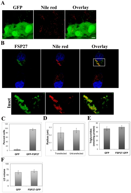
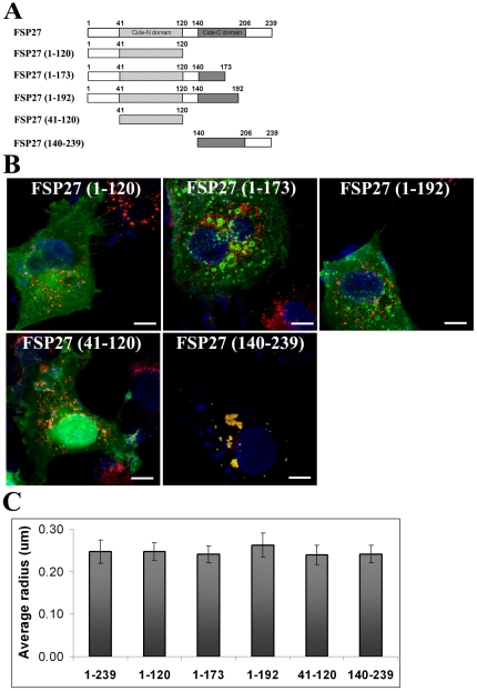
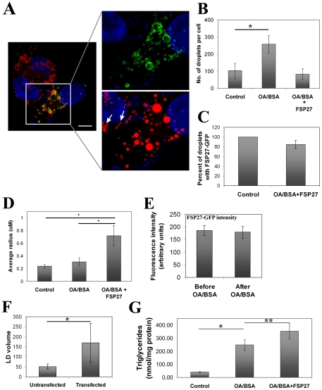
Fat Specific Protein 27 (FSP27), a lipid droplet (LD) associated protein in adipocytes, regulates triglyceride (TG) storage. In the present study we demonstrate that FSP27 plays a key role in LD morphology to accumulate TGs. We show here that FSP27 promotes clustering of the LDs which is followed by their fusion into fewer and enlarged droplets. To map the domains of FSP27 responsible for these events, we generated GFP-fusion constructs of deletion mutants of FSP27. Microscopic analysis revealed that amino acids 173-220 of FSP27 are necessary and sufficient for both the targeting of FSP27 to LDs and the initial clustering of the droplets. Amino acids 120-140 are essential but not sufficient for LD enlargement, whereas amino acids 120-210 are necessary and sufficient for both clustering and fusion of LDs to form enlarged droplets. In addition, we found that FSP27-mediated enlargement of LDs, but not their clustering, is associated with triglyceride accumulation. These results suggest a model in which FSP27 facilitates LD clustering and then promotes their fusion to form enlarged droplets in two discrete, sequential steps, and a subsequent triglyceride accumulation.


Conflict of interest statement

Competing Interests: The authors have declared that no competing interests exist.

Figures

Figure 1
Figure 1. FSP27-GFP expression causes clustering of LDs in COS-7 cells.
A, COS-7 cells were transfected with GFP vector alone. Left panel shows GFP expression in cells after 16 hr of transfection. The LDs stained with Nile red are shown in red color in the middle panel. Bar, 10 µm. B, COS-7 cells transfected with FSP27-GFP for 16 hr. Nucleus was stained with DAPI. Left panel shows cell expressing FSP27-GFP (top) and non-expressing cell (bottom) in the same fied of the microscope. The middle panel shows LDs stained with Nile red. A 2 µm Z-slice of the transfected cell (in the inset of top right panel) is enlarged in the lower panels to show a clear representation of FSS7-GFP distribution around the clustered droplets (also see Figure S1). Bar, 10 µm. C, percent of GFP or FSP27-GFP expressing COS-7 cells which showed clustered LDs. Results are an average of at least three independent experiments ± standard deviation. About 25 cells were counted in each individual experiment (p<0.0001). D, morphometric analysis was performed to measure the radius of LDs in FSP27-GFP transfected and untransfected COS-7 cells. Results are an average of at least 25 cells from three independent experiments ± standard deviation. E, Biochemical quantification of total triglycerides in COS-7 cells after 16 hr of transfection with GFP and FSP27-GFP cDNA. Data is from three independent experiments, error bars show standard deviations (p = 0.17). F, total LD volume in cells transfected with GFP vector alone and FSP27-GFP. The bars indicate the volume in µm3 ± standard error, (*, p = 0.3).
Figure 2
Figure 2. Carboxy-terminus of FSP27 is necessary for LD targeting and clustering.
A, deletion mutations of FSP27. GFP fusion constructs of these deletion mutations were prepared to identify the LD targeting and clustering regions of FSP27. B, expression of GFP fusion constructs of FSP27 deletion mutants in COS-7 cells. The images show the distribution of mutants (green) after 16 hr of transfection. LDs were labeled with Nile red (red) and nucleus was labeled with DAPI (blue). C, morphometric analysis was performed on microscopic images to measure the radius of LDs. The results shown are an average of mean radius of LDs in at least 10 cells from three different experiments in each condition. The error bars show standard deviation.
Figure 3
Figure 3. Amino acids 173–220 are necessary and sufficient for localization of FSP27 to LDs and their clustering.
A, deletion mutants in the C-terminus of FSP27 which were fused with GFP to identify the minimum essential domain of FSP27 required for its targeting to LDs and cluster them. B, expression of GFP fusion constructs of FSP27 deletion mutants in COS-7 cells. The images show the distribution of mutants (green) after 16 hr of transfection. LDs were labeled with Nile red (red) and nucleus was labeled with DAPI (blue). C, amino acids 173–220 of FSP27 are necessary and sufficient for targeting to the LDs and clustering them. Left panel shows FSP27 (173–220)-GFP expression after 16 hr of transfection, middle panel shows the LDs labeled with Nile red. Bar 10 µm. D, morphometric analysis was performed on microscopic images to measure the radius of LDs. The results shown are an average of mean radius of LDs in at least 10 cells from three different experiments in each condition. The error bars show standard deviation.
Figure 4
Figure 4. Clustering of LDs is followed by their enlargement in FSP27-GFP expressing COS-7 cells.
A, time course of FSP27-GFP expression in COS-7 cells. COS-7 cells were transfected with FSP27-GFP. After 4 hr, 16 hr and 24 hr of transfection, the cells were fixed, labeled with Nile red and analyzed under confocal microscope. After 4 hr of FSP27 expression almost all the LDs were associated with FSP27 (left panel), at 16 hr the LDs in about 85% of FSP27-GFP expressing cells were clustered (middle panel) and at 24 hr after FSP27 transfection about 80–85% of the cells had enlarged LDs (right panel). Bar 10 µm. B, Morphometric analysis was performed on microscopic images to measure the average radius of LDs. The results are from at least 10 cells in each condition form three independent experiments. The error bars show standard deviation, (*, p<0.001). C, Quantification of LDs per cell was performed in GFP and FSP27-GFP expressing cells at different time points. The results are an average of LDs per cell. LDs in at least 10 cells were counted in each condition from three independent experiments, (*, p<0.001). D, biochemical quantification of total TGs in COS-7 cells at different time points after transfection with GFP and FSP27-GFP. Data is from three independent experiments, error bars show standard deviation, (*, p<0.001).
Figure 5
Figure 5. Amino acids 120–210 are necessary and sufficient for enlargement of LDs.
A, deletion mutants of FSP27 which were fused with GFP to identify the minimum essential domain of FSP27 associated with LD enlargement. B, expression of GFP fusion constructs of FSP27 deletion mutants in COS-7 cells. The images show the distribution of mutants (green) after 16 hr of transfection. LDs were labeled with Nile red (red) and nucleus was labeled with DAPI (blue). Bar 10 µm. C, morphometric analysis was performed on microscopic images to measure the radius of LDs. The results shown are an average of mean radius of LDs in at least 10 cells from three different experiments in each condition. The error bars show standard deviation.
Figure 6
Figure 6. FSP27 (120–210) rapidly clusters and enlarges the LDs.
A, LD morphology after 24 hr of transfecting FSP27 (173–220), FSP27 (120–210), and full length FSP27 in COS-7 cells. LDs were stained with Nile red. Bar 10 µm. B, COS-7 cells were transfected with FSP27 (120–210) and analyzed after 4 hr and 8 hr of transfection. LDs were stained with Nile red. Bar 10 µm.
Figure 7
Figure 7. FSP27 redistributes to newly formed LDs and might promote coalescence of droplets to form enlarged LDs.
A, COS-7 cells were transfected with FSP27-GFP, 4 hr after transfection 70 µM cycloheximide was added for 1 hr to inhibit protein synthesis. The cells were then fed with 60 µM OA/BSA for the formation of new LDs overnight during which 70 µM cycloheximide was also present. After overnight incubation the cells were washed, incubated for another 1 hr in the absence of OA/BSA and presence of cycloheximide, fixed and stained with Nile red. Note that the FSP27-GFP expressing cells have fewer but enlarged LDs and FSP27-GFP was associated with most of the droplets independent of their size. In contrast, the neighboring cells that do not express FSP27-GFP, are filled with large numbers of small LDs and all of these droplets are of similar size, also, in spite of close packing of the droplets (shown by arrows in the right bottom image) they do not show any sign of coalescence. B, quantification of number of droplets per cell under conditions mentioned above. In control, the cells were fed with OA/BSA alone. (*, p<0.0001). C, percent of droplets associated with FSP27-GFP after overnight OA/BSA treatment of COS-7 cells. D, average radius of the lipid droplets. In control, the cells were fed with BSA alone (*, p<0.0001). E, fluorescence intensity of FSP27-GFP before and after OA/BSA feeding in the presence of cycloheximide under the conditions mentioned in panel A. F, total LD volume in untransfected and FSP27-GFP transfected cells fed with OA/BSA. The bars indicate the volume in µm3 ± standard deviation. (*, p<0.002). G, biochemical analysis of TG content in COS-7 cells. The cells transfected with GFP had 5.5-fold increase in TGs when fed with OA/BSA (*, p<0.0001). The cells transfected with FSP27-GFP and fed with OA/BSA showed further increase in TG content (**, p<0.005). In B–F, the cells were treated with cycloheximide and fed with OA/BSA in the exact same conditions mentioned in panel A.
Figure 8
Figure 8. Schematic representation of FSP27 and its functional domains.
Two domains of FSP27 have been reported in literature, CIDE-N and CIDE-C. The CIDE-N and CIDE-C domain ranges from amino acids 41–120 and 140–206 respectively . The N-terminus of FSP27 is from amino acids 1–120 and the C-terminus is from amino acids 121–239. We found that FSP27-mediated enlargement of LDs consists of two independent steps, clustering followed by fusion of LDs. Amino acids 172–210 are necessary and sufficient for FSP27-mediated clustering of LDs. The clustering of LDs has no effect on their size and cellular TG levels. The LD clustering is followed by their enlargement. Amino acids 120–210 are sufficient for clustering and enlargement of LDs. Interestingly, FSP27-mediated enlargement of LDs is accompanied with increased TG levels in the cells. These results highlight the important role of FSP27 in LD morphology and TG accumulation.

References

    1. Beckman M. Cell biology. Great balls of fat. Science. 2006;311:1232–1234. - PubMed
    1. Farese RV, Walther TC. Lipid droplets finally get a little R-E-S-P-E-C-T. Cell. 2009;139:855–860. - PMC - PubMed
    1. Brasaemle DL. Thematic review series: adipocyte biology. The perilipin family of structural lipid droplet proteins: stabilization of lipid droplets and control of lipolysis. J Lipid Res. 2007;48:2547–2559. - PubMed
    1. Ducharme NA, Bickel PE. Lipid droplets in lipogenesis and lipolysis. Endocrinology. 2008;149:942–949. - PubMed
    1. Wolins NE, Brasaemle DL, Bickel PE. A proposed model of fat packaging by exchangeable lipid droplet proteins. FEBS Lett. 2006;580:5484–5491. - PubMed

Publication types