. 2011;6(12):e28717.
doi: 10.1371/journal.pone.0028717. Epub 2011 Dec 14.

Ursodeoxycholic acid is conjugated with taurine to promote secretin-stimulated biliary hydrocholeresis in the normal rat

Affiliations

Ursodeoxycholic acid is conjugated with taurine to promote secretin-stimulated biliary hydrocholeresis in the normal rat

Miriam Úriz et al. PLoS One. 2011.

Abstract

Background & aims: Secretin induces bicarbonate-rich hydrocholeresis in healthy individuals, but not in untreated patients with primary biliary cirrhosis (PBC). Ursodeoxycholic acid (UDCA)--the first choice treatment for PBC--restores the secretin response. Compared with humans, secretin has poor effect in experimental normal-rat models with biliary drainage, although it may elicit hydrocholeresis when the bile-acid pool is maintained. In view of the benefits of UDCA in PBC, we used normal-rat models to unravel the acute contribution of UDCA (and/or taurine-conjugated TUDCA) for eliciting the biliary secretin response.

Methods: Intravascular and/or intrabiliary administration of agonists and inhibitors was performed in normal rats with biliary monitoring. Secretin/bile-acid interplay was analyzed in 3D cultured rat cholangiocytes that formed expansive cystic structures with intralumenal hydroionic secretion.

Results: In vivo, secretin stimulates hydrocholeresis upon UDCA/TUDCA infusion, but does not modify the intrinsic hypercholeretic effect of dehydrocholic acid (DHCA). The former effect is dependent on microtubule polymerization, and involves PKCα, PI3K and MEK pathways, as shown by colchicine (i.p.) and retrograde biliary inhibitors. In vitro, while secretin alone accelerates the spontaneous expansion of 3D-cystic structures, this effect is enhanced in the presence of TUDCA, but not UDCA or DHCA. Experiments with inhibitors and Ca(2+)-chelator confirmed that the synergistic effect of secretin plus TUDCA involves microtubules, intracellular Ca(2+), PKCα, PI3K, PKA and MEK pathways. Gene silencing also demonstrated the involvement of the bicarbonate extruder Ae2.

Conclusions: UDCA is conjugated in order to promote secretin-stimulated hydrocholeresis in rats through Ae2, microtubules, intracellular Ca(2+), PKCα, PI3K, PKA, and MEK.


Conflict of interest statement

Competing Interests: The authors have declared that no competing interests exist.

Figures

Figure 1
Figure 1. Secretin stimulates bile flow in normal rats infused with UDCA and TUDCA.
The choleretic effects of secretin, UDCA, or TUDCA administered alone were not significant, while infusion of DHCA alone stimulated the bile flow of normal rats, with no effect on the secretin response. All relevant differences are indicated (with no statistical difference between secretin-stimulated bile flows in UDCA- and TUDCA-infused animals). Data are shown as mean ± SD.
Figure 2
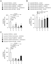
Figure 2. Increased bile flow in rats receiving secretin and either UDCA or TUDCA is dependent on microtubule polymerization, PKC, PI3K and MEK.
The increased bile flow in rats receiving secretin plus (A) UDCA or (B) TUDCA was blocked with intraperitoneal administration of colchicine (an inhibitor of microtubule polymerization), intrabiliary administration of the Ca2+-dependent PKCα inhibitor Gö6976, intrabiliar wortmannin (a PI3K inhibitor), and intrabiliar U0126 (a MEK inhibitor). (C) On the other hand, the hydrocholeretic effect to DHCA was unaffected by these inhibitors. Increases versus respective controls were calculated as indicated. Data are shown as mean ± SD.
Figure 3
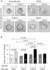
Figure 3. TUDCA promotes secretin-stimulated expansion of 3D-cultured cholangiocyte cystic structures.
(A) Representative images of cystic structures incubated for 60 min in the presence or absence of bile acids (UDCA, TUDCA, or DHCA), and with secretin for the last 30 min. (B) While secretin alone stimulated cystic expansion, such an expansion was further accelerated in the presence of TUDCA, but not in the presence of UDCA or DHCA (which rather tended to block the stimulatory effect of secretin). Only comparisons of interest are indicated.
Figure 4
Figure 4. The expansion of 3D-cultured cholangiocyte cystic structures stimulated by the combination of secretin and TUDCA is dependent on microtubule polymerization, intracellular Ca2+ and PKCα signaling, and PI3K, PKA and MEK pathways.
[TUDCA+secretin]-stimulated cystic expansion was sensitive to colchicine, the Ca2 +-chelator BAPTA, Gö6976, wortmannin, U0126 and the PKA-inhibitor H89. Cystic expansion is expressed as percentage of the area at the end of the experiment (60 min) versus initial area (at time 0). Data are shown as mean ± SEM.
Figure 5
Figure 5. The bicarbonate extruder Ae2 is involved in the concerted hydrocholeretic effect of TUDCA and secretin.
While the presence of control siRNA had no effect on the expansion rate of 3D-cultured cholangiocyte cystic structures stimulated by secretin alone or combination of secretin and TUDCA (left), the specific rAe2 siRNA against rat Ae2 mRNA blocked all stimulatory effects (right). Data are shown as mean ± SEM.
Figure 6
Figure 6. Mechanisms proposed for the TUDCA-mediated secretin-stimulated bile flow in the normal rat.
The diagrams summarize the proposed mechanisms for the concerted action of UDCA and secretin that stimulates further the hydrocholeresis in the normal-rat biliary epithelium: UDCA is conjugated with taurine in the hepatocytes and secreted to bile as TUDCA, which may directly access the apical side of cholangiocytes (as well as the basolateral side in the case of an intact animal with enterohepatic circulation). In cholangiocytes, TUDCA may favor the action of secretin through modulation of intracellular Ca2 + levels, and PKCα and PI3K activation. MEK and cAMP-dependent pathways as well as mobilization of vesicles appear to be also involved. Ae2 has a crucial role for the secretin-stimulated bicarbonate-rich hydrocholeresis by acting as the ultimate extruder of bicarbonate through Cl/HCO3 exchange. Neither DHCA nor unconjugate UDCA may exert any of these acute effects on the biliary epithelium, and they promote hydrocholeresis via different mechanisms.

References

    1. Banales JM, Prieto J, Medina JF. Cholangiocyte anion exchange and biliary bicarbonate excretion. World J Gastroenterol. 2006;12:3496–3511. - PMC - PubMed
    1. Kanno N, LeSage G, Glaser S, Alpini G. Regulation of cholangiocyte bicarbonate secretion. Am J Physiol Gastrointest Liver Physiol. 2001;281:G612–625. - PubMed
    1. Banales JM, Arenas F, Rodríguez-Ortigosa CM, Sáez E, Uriarte I, et al. Bicarbonate-rich choleresis induced by secretin in normal rat is taurocholate-dependent and involves AE2 anion exchanger. Hepatology. 2006;43:266–275. - PubMed
    1. Tietz PS, Marinelli RA, Chen XM, Huang B, Cohn J, et al. Agonist-induced coordinated trafficking of functionally related transport proteins for water and ions in cholangiocytes. J Biol Chem. 2003;278:20413–20419. - PubMed
    1. Poupon R. Primary biliary cirrhosis: a 2010 update. J Hepatol. 2010;52:745–758. - PubMed

Publication types

MeSH terms