

The distribution of Toxoplasma gondii cysts in the brain of a mouse with latent toxoplasmosis: implications for the behavioral manipulation hypothesis
- PMID: 22194951
- PMCID: PMC3237564
- DOI: 10.1371/journal.pone.0028925
The distribution of Toxoplasma gondii cysts in the brain of a mouse with latent toxoplasmosis: implications for the behavioral manipulation hypothesis
Abstract
Background: The highly prevalent parasite Toxoplasma gondii reportedly manipulates rodent behavior to enhance the likelihood of transmission to its definitive cat host. The proximate mechanisms underlying this adaptive manipulation remain largely unclear, though a growing body of evidence suggests that the parasite-entrained dysregulation of dopamine metabolism plays a central role. Paradoxically, the distribution of the parasite in the brain has received only scant attention.
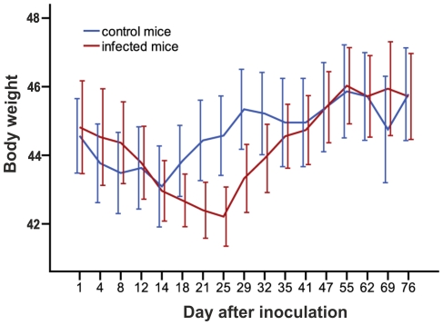
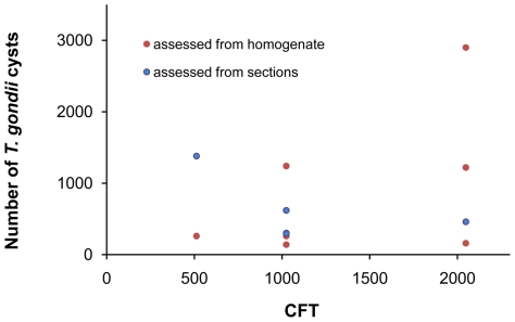
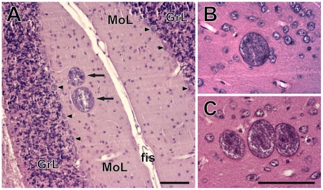
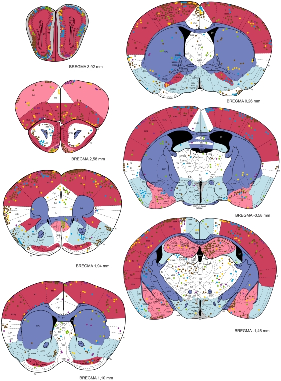
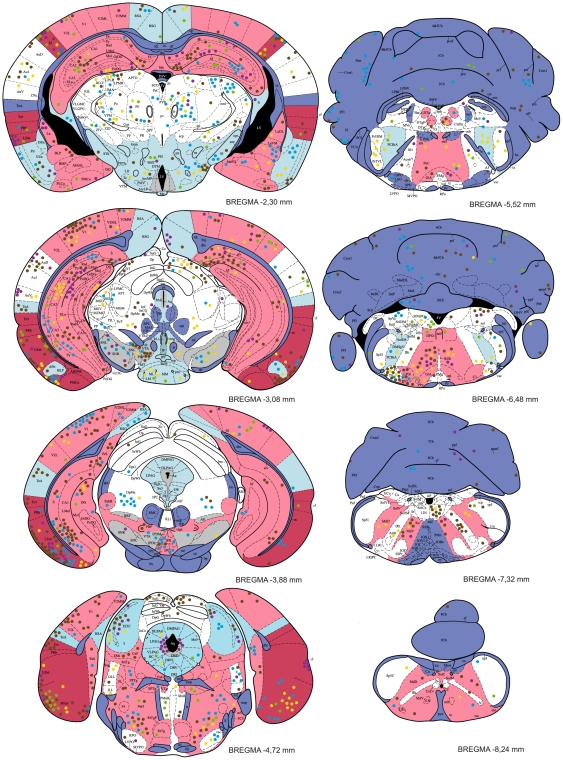
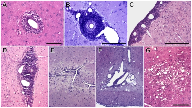
Methodology/principal findings: The distributions of T. gondii cysts and histopathological lesions in the brains of CD1 mice with latent toxoplasmosis were analyzed using standard histological techniques. Mice were infected per orally with 10 tissue cysts of the avirulent HIF strain of T. gondii at six months of age and examined 18 weeks later. The cysts were distributed throughout the brain and selective tropism of the parasite toward a particular functional system was not observed. Importantly, the cysts were not preferentially associated with the dopaminergic system and absent from the hypothalamic defensive system. The striking interindividual differences in the total parasite load and cyst distribution indicate a probabilistic nature of brain infestation. Still, some brain regions were consistently more infected than others. These included the olfactory bulb, the entorhinal, somatosensory, motor and orbital, frontal association and visual cortices, and, importantly, the hippocampus and the amygdala. By contrast, a consistently low incidence of tissue cysts was recorded in the cerebellum, the pontine nuclei, the caudate putamen and virtually all compact masses of myelinated axons. Numerous perivascular and leptomeningeal infiltrations of inflammatory cells were observed, but they were not associated with intracellular cysts.
Conclusion/significance: The observed pattern of T. gondii distribution stems from uneven brain colonization during acute infection and explains numerous behavioral abnormalities observed in the chronically infected rodents. Thus, the parasite can effectively change behavioral phenotype of infected hosts despite the absence of well targeted tropism.
Conflict of interest statement
Figures
References
-
- Hutchison WM, Work K. Toxoplasma – a versatile parasite. New Sci. 1969;29:464–466.
-
- Webster JP. Rats, cats, people and parasites: the impact of latent toxoplasmosis on behaviour. Microbes Infect. 2001;3:1037–1045. - PubMed
-
- Flegr J. Influence of latent toxoplasmosis on the phenotype of intermediate hosts. Folia Parasitol (Praha) 2010;57:81–7. - PubMed
-
- Vyas A, Sapolsky R. Manipulation of host behaviour by Toxoplasma gondii: what is the minimum a proposed proximate mechanism should explain? Folia Parasitol (Praha) 2010;57:88–94. - PubMed
Publication types
MeSH terms
Substances
LinkOut - more resources
Full Text Sources
Medical
Miscellaneous
