. 2011;6(12):e28970.
doi: 10.1371/journal.pone.0028970. Epub 2011 Dec 14.

Variation in mesoderm specification across Drosophilids is compensated by different rates of myoblast fusion during body wall musculature development

Affiliations

Variation in mesoderm specification across Drosophilids is compensated by different rates of myoblast fusion during body wall musculature development

Mirela Belu et al. PLoS One. 2011.

Abstract

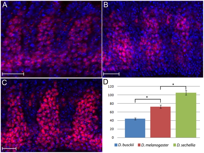
Background: It has been shown that species separated by relatively short evolutionary distances may have extreme variations in egg size and shape. Those variations are expected to modify the polarized morphogenetic gradients that pattern the dorso-ventral axis of embryos. Currently, little is known about the effects of scaling over the embryonic architecture of organisms. We began examining this problem by asking if changes in embryo size in closely related species of Drosophila modify all three dorso-ventral germ layers or only particular layers, and whether or not tissue patterning would be affected at later stages.

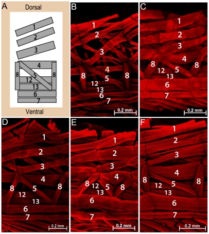
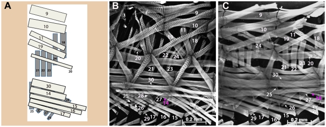
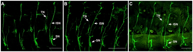
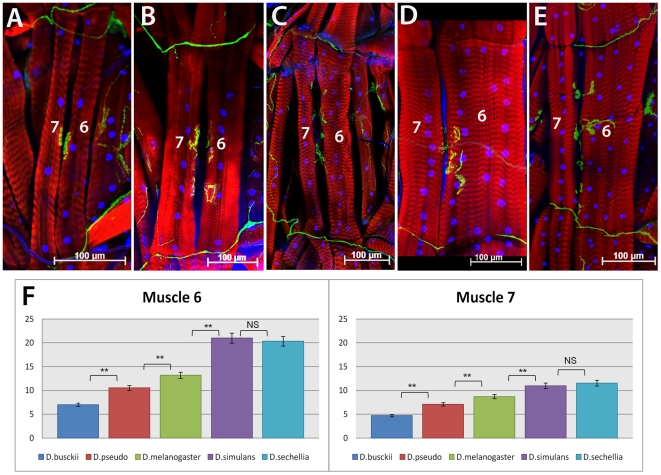
Principal findings: Here we report that changes in scale affect predominantly the mesodermal layer at early stages, while the neuroectoderm remains constant across the species studied. Next, we examined the fate of somatic myoblast precursor cells that derive from the mesoderm to test whether the assembly of the larval body wall musculature would be affected by the variation in mesoderm specification. Our results show that in all four species analyzed, the stereotyped organization of the body wall musculature is not disrupted and remains the same as in D. melanogaster. Instead, the excess or shortage of myoblast precursors is compensated by the formation of individual muscle fibers containing more or less fused myoblasts.

Conclusions: Our data suggest that changes in embryonic scaling often lead to expansions or retractions of the mesodermal domain across Drosophila species. At later stages, two compensatory cellular mechanisms assure the formation of a highly stereotyped larval somatic musculature: an invariable selection of 30 muscle founder cells per hemisegment, which seed the formation of a complete array of muscle fibers, and a variable rate in myoblast fusion that modifies the number of myoblasts that fuse to individual muscle fibers.


Conflict of interest statement

Competing Interests: The authors have declared that no competing interests exist.

Figures

Figure 1
Figure 1. Mesodermal domain varies with embryo size.
A–C) Cross-section of blastoderm stage embryos stained for mesodermal marker snail (sna, red) and nuclear dye Hoescht (green). A) D. busckii; B) D. melanogaster, and C) D. sechellia. Width of presumptive mesoderm is indicated by arrowheads and contains about 14 nuclei expression snail for D. busckii (A), 19 nuclei for D. melanogaster (B) and 26 for D. sechellia (C). Scale bar: 100 µm. D–I) Ventral view of whole mounted embryos adjusted to same size. sna is labeled in red, nuclei are labeled in green. D) D. busckii whole embryo and (E) high magnification detail of cells labeled with sna (left, sna and stained nuclei; right, nuclei only). F, G) D. melanogaster; H, I) D. sechellia. Brackets indicate extension of sna+ nuclei.
Figure 2
Figure 2. Expression pattern of Dmef 2 reveals differences in somatic myoblast numbers between Drosophila species.
Lateral view of whole mounted mid-E12 stage embryos stained for anti-DMef2 antibody (red) and Hoescht (blue). External most layer of D-Mef2 positive cells are shown for (A) D. busckii, (B) D. melanogaster and (C) D. sechellia. D) The numbers of D-Mef2 positive cells are decreased to an average of 44 in D. busckii and increased in D. sechellia to an average of 105, in comparison to D. melanogaster, which has an average of 72 cells. Anterior to the left, dorsal is up. Scale bar: 20 µm. Sample size, n = 5 hemisegments. Asterisks indicate p-values of p = 0.0057 and p = 0.0059.
Figure 3
Figure 3. The pattern of internal muscle layer is identical in all species analyzed.
A) Schematic representation of the larval body wall depicting the internal muscle layer of one abdominal hemisegment, as previously described for D. melanogaster (adapted from Bate, M., 1990 and 1993). Ventral and dorsal positions indicated in (A) also correspond to orientation of images shown in (BF). Anterior is to the left. Dissected L3 larva of B) D. busckii; C) D. pseudoobscura; D) D. melanogaster; E) D. simulans and (F) D. sechellia species. The muscle fibers were stained with phalloidin (red). Each internal muscle fiber (indicated by its corresponding number) is present in all species, and displays similar orientation and attachment site as the stereotyped pattern described for D. melanogaster (A, D). Scale bars: 0.2 mm.
Figure 4
Figure 4. Conservation of external and intermediate body wall muscle layer in L3 larva.
A) Scheme of external (dark gray) and intermediate (light gray) abdominal muscle layers of one hemisegment, as previously described for D. melanogaster (adapted from Bate, M., 1990 and 1993). B) D. busckii; C) D. sechellia. Both the external and intermediate muscle layers (grayscale) are indicated by numbers in two Drosophila species: D. busckii (B), and D. sechellia (C). Two adjacent hemisegments are shown in (B–C) to allow better visualization of each external and intermediate muscle fibers. Ventral, dorsal and anterior positions indicated in (A) also correspond to orientation of images shown in (BC). Scale bars: 0.2 mm.
Figure 5
Figure 5. Peripheral motor nerves display stereotyped trajectories and innervation points at late embryonic stages.
Anti-Fas II staining in E16–17 embryos from (A) D. melanogaster; B) D. busckii; and (C) D. sechellia. Major peripheral nerves indicated by ISN, SN, and TN have similar organization and branch at similar positions in all species analyzed. Lateral view of dissected embryo fillets, anterior to left, dorsal is up. Scale bar: 100 µm.
Figure 6
Figure 6. Variation in number of nuclei for muscle fibers 6 and 7 in Drosophilid larva.
A) D. busckii; B) D. pseudoobscura; C) D. melanogaster; D) D. simulans and (E) D. sechellia abdominal muscle fibers 6 and 7 in L3 stage larva. The muscle fibers were stained with phalloidin (red), neuromuscular junctions with anti-HRP (green) and nuclei with Hoechst (blue). D. busckii and D. pseudoobscura (A, B) have muscle 6 and 7 reduced in size compared to D. melanogaster (C), while D. simulans and D. sechellia (D, E) have the largest fibers of all species analyzed. F) Quantification graph of nuclei counts per fiber 6 and 7. Species are indicated in legend. NS, “no statistically significant difference”. Asterisks ** indicate a p value<0.001 (See text for exact values). Scale bars: 100 µm. Anterior side is up.

References

    1. Gregor T, Bialek W, de Ruyter van Steveninck RR, Tank DW, Wieschaus EF. Diffusion and scaling during early embryonic pattern formation. Proc Natl Acad Sci U S A. 2005;102:18403–18407. - PMC - PubMed
    1. Lott SE, Kreitman M, Palsson A, Alekseeva E, Ludwig MZ. Canalization of segmentation and its evolution in Drosophila. Proc Natl Acad Sci U S A. 2007;104:10926–10931. - PMC - PubMed
    1. Markow TA, Beall S, Matzkin LM. Egg size, embryonic development time and ovoviviparity in Drosophila species. J Evol Biol. 2009;22:430–434. - PubMed
    1. Irish VF, Gelbart WM. The decapentaplegic gene is required for dorsal-ventral patterning of the Drosophila embryo. Genes Dev. 1987;1:868–879. - PubMed
    1. Roth S, Stein D, Nusslein-Volhard C. A gradient of nuclear localization of the dorsal protein determines dorsoventral pattern in the Drosophila embryo. Cell. 1989;59:1189–1202. - PubMed

Publication types

MeSH terms

Substances

LinkOut - more resources