Synchrotron radiation X-ray microfluorescence reveals polarized distribution of atomic elements during differentiation of pluripotent stem cells
- PMID: 22195032
- PMCID: PMC3241705
- DOI: 10.1371/journal.pone.0029244
Synchrotron radiation X-ray microfluorescence reveals polarized distribution of atomic elements during differentiation of pluripotent stem cells
Abstract
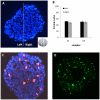
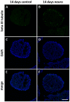
The mechanisms underlying pluripotency and differentiation in embryonic and reprogrammed stem cells are unclear. In this work, we characterized the pluripotent state towards neural differentiated state through analysis of trace elements distribution using the Synchrotron Radiation X-ray Fluorescence Spectroscopy. Naive and neural-stimulated embryoid bodies (EB) derived from embryonic and induced pluripotent stem (ES and iPS) cells were irradiated with a spatial resolution of 20 µm to make elemental maps and qualitative chemical analyses. Results show that these embryo-like aggregates exhibit self-organization at the atomic level. Metallic elements content rises and consistent elemental polarization pattern of P and S in both mouse and human pluripotent stem cells were observed, indicating that neural differentiation and elemental polarization are strongly correlated.
Conflict of interest statement
Figures


Similar articles
-
Stable expression of FoxA1 promotes pluripotent P19 embryonal carcinoma cells to be neural stem-like cells.Gene Expr. 2012;15(4):153-62. doi: 10.3727/105221612x13372578119571. Gene Expr. 2012. PMID: 22783724 Free PMC article.
-
Mouse embryonic stem cells irradiated with γ-rays differentiate into cardiomyocytes but with altered contractile properties.Mutat Res. 2013 Aug 30;756(1-2):37-45. doi: 10.1016/j.mrgentox.2013.06.007. Epub 2013 Jun 20. Mutat Res. 2013. PMID: 23792212
-
Neural differentiation of human embryonic stem cells as an in vitro tool for the study of the expression patterns of the neuronal cytoskeleton during neurogenesis.Biochem Biophys Res Commun. 2013 Sep 13;439(1):154-9. doi: 10.1016/j.bbrc.2013.07.130. Epub 2013 Aug 11. Biochem Biophys Res Commun. 2013. PMID: 23939048
-
Roles of TGF-β family signals in the fate determination of pluripotent stem cells.Semin Cell Dev Biol. 2014 Aug;32:98-106. doi: 10.1016/j.semcdb.2014.05.017. Epub 2014 Jun 6. Semin Cell Dev Biol. 2014. PMID: 24910449 Review.
-
Pluripotent Stem Cells and DNA Damage Response to Ionizing Radiations.Radiat Res. 2016 Jul;186(1):17-26. doi: 10.1667/RR14417.1. Epub 2016 Jun 22. Radiat Res. 2016. PMID: 27332952 Free PMC article. Review.
Cited by
-
Potassium as a pluripotency-associated element identified through inorganic element profiling in human pluripotent stem cells.Sci Rep. 2017 Jul 10;7(1):5005. doi: 10.1038/s41598-017-05117-2. Sci Rep. 2017. PMID: 28694442 Free PMC article.
-
Trace elements during primordial plexiform network formation in human cerebral organoids.PeerJ. 2017 Feb 8;5:e2927. doi: 10.7717/peerj.2927. eCollection 2017. PeerJ. 2017. PMID: 28194309 Free PMC article.
-
Sub-micrometer scale synchrotron x-ray fluorescence measurements of trace elements in teeth compared with laser ablation inductively coupled plasma mass spectrometry.J Expo Sci Environ Epidemiol. 2025 Jul;35(4):625-629. doi: 10.1038/s41370-025-00754-6. Epub 2025 Jan 29. J Expo Sci Environ Epidemiol. 2025. PMID: 39881199 Free PMC article.
-
A unique feature of iron loss via close adhesion of Helicobacter pylori to host erythrocytes.PLoS One. 2012;7(11):e50314. doi: 10.1371/journal.pone.0050314. Epub 2012 Nov 21. PLoS One. 2012. PMID: 23185604 Free PMC article.
-
Using X-ray in-line phase-contrast imaging for the investigation of nude mouse hepatic tumors.PLoS One. 2012;7(6):e39936. doi: 10.1371/journal.pone.0039936. Epub 2012 Jun 28. PLoS One. 2012. PMID: 22761929 Free PMC article.
References
-
- Smith A. The battlefield of pluripotency. Cell. 2005;123:757–760. - PubMed
-
- Takahashi K, Yamanaka S. Induction of pluripotent stem cells from mouse embryonic and adult fibroblast cultures by defined factors. Cell. 2006;126:663–676. - PubMed
-
- Chan EM, Ratanasirintrawoot S, Park IH, Manos PD, Loh YH, et al. Live cell imaging distinguishes bona fide human iPS cells from partially reprogrammed cells. Nat Biotechnol. 2009;27:1033–1037. - PubMed
-
- Qin J, Guo X, Cui GH, Zhou YC, Zhou DR, et al. Cluster characterization of mouse embryonic stem cell-derived pluripotent embryoid bodies in four distinct developmental stages. Biologicals. 2009;37:235–244. - PubMed
Publication types
MeSH terms
Substances
LinkOut - more resources
Full Text Sources

