. 2011;6(12):e29246.
doi: 10.1371/journal.pone.0029246. Epub 2011 Dec 15.

Sca-1+ cardiac stem cells mediate acute cardioprotection via paracrine factor SDF-1 following myocardial ischemia/reperfusion

Affiliations

Sca-1+ cardiac stem cells mediate acute cardioprotection via paracrine factor SDF-1 following myocardial ischemia/reperfusion

Chunyan Huang et al. PLoS One. 2011.

Abstract

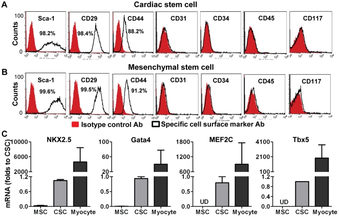
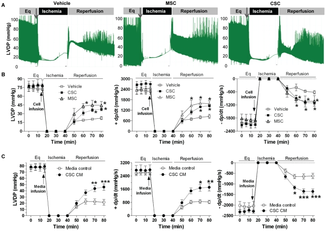
Background: Cardiac stem cells (CSCs) promote myocardial recovery following ischemia through their regenerative properties. However, little is known regarding the implication of paracrine action by CSCs in the setting of myocardial ischemia/reperfusion (I/R) injury although it is well documented that non-cardiac stem cells mediate cardioprotection via the production of paracrine protective factors. Here, we studied whether CSCs could initiate acute protection following global myocardial I/R via paracrine effect and what component from CSCs is critical to this protection.

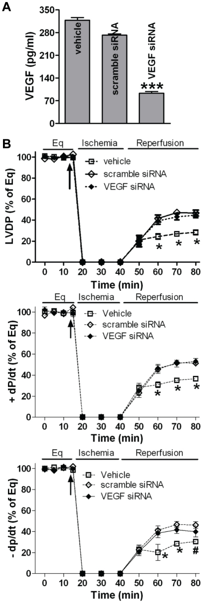
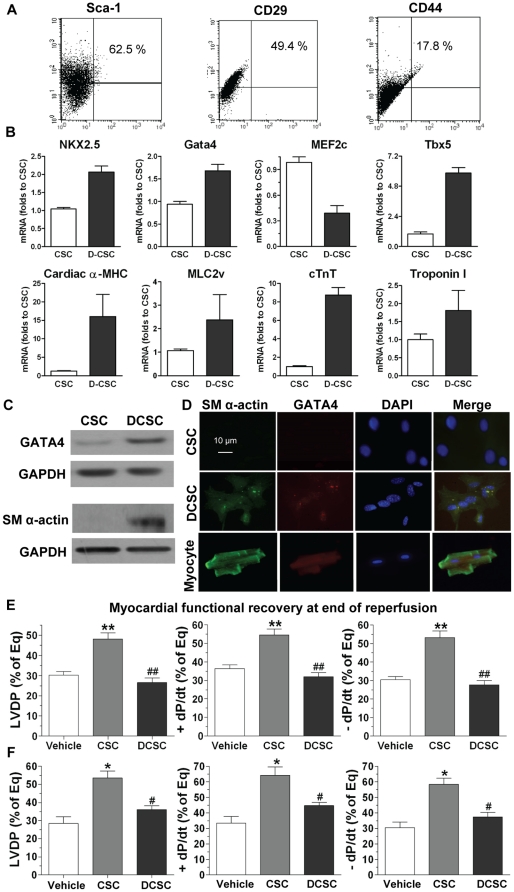
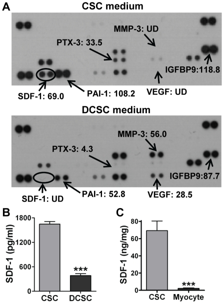
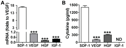
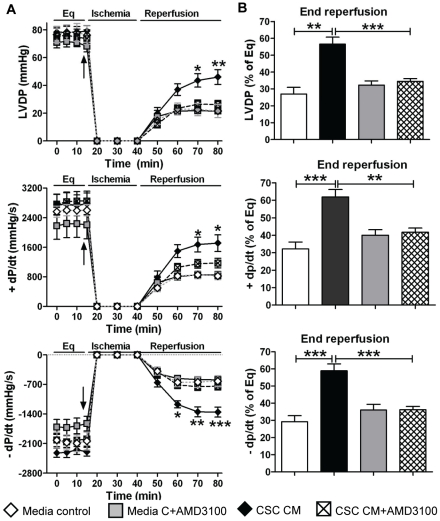
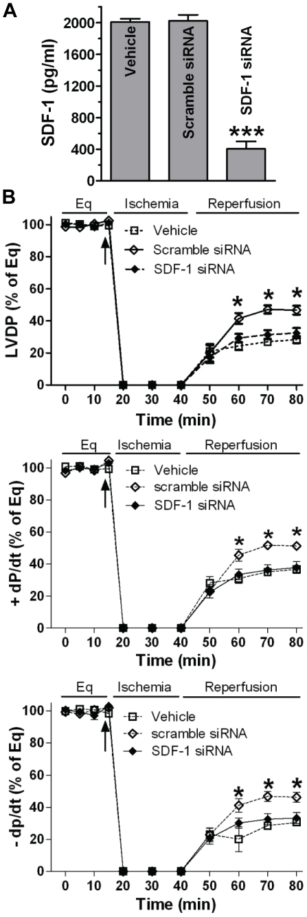
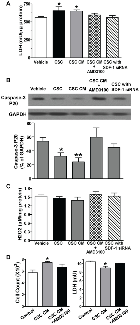
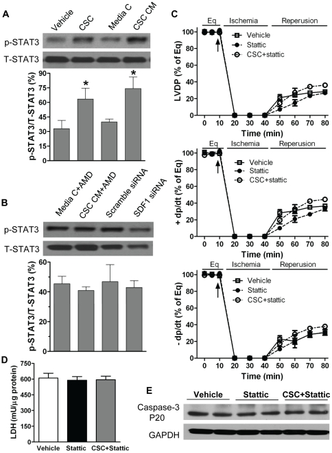
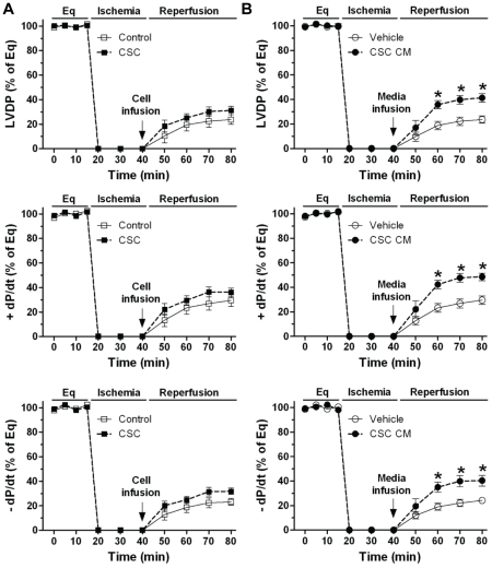
Methodology/principal findings: A murine model of global myocardial I/R was utilized to investigate paracrine effect of Sca-1+ CSCs on cardiac function. Intracoronary delivery of CSCs or CSC conditioned medium (CSC CM) prior to ischemia significantly improved myocardial function following I/R. siRNA targeting of VEGF in CSCs did not affect CSC-preserved myocardial function in response to I/R injury. However, differentiation of CSCs to cardiomyocytes (DCSCs) abolished this protection. Through direct comparison of the protein expression profiles of CSCs and DCSCs, SDF-1 was identified as one of the dominant paracrine factors secreted by CSCs. Blockade of the SDF-1 receptor by AMD3100 or downregulated SDF-1 expression in CSCs by specific SDF-1 siRNA dramatically impaired CSC-induced improvement in cardiac function and increased myocardial damage following I/R. Of note, CSC treatment increased myocardial STAT3 activation after I/R, whereas downregulation of SDF-1 action by blockade of the SDF-1 receptor or SDF-1 siRNA transfection abolished CSC-induced STAT3 activation. In addition, inhibition of STAT3 activation attenuated CSC-mediated cardioprotection following I/R. Finally, post-ischemic infusion of CSC CM was shown to significantly protect I/R-caused myocardial dysfunction.

Conclusions/significance: This study suggests that CSCs acutely improve post-ischemic myocardial function through paracrine factor SDF-1 and up-regulated myocardial STAT3 activation.


Conflict of interest statement

Competing Interests: The authors have declared that no competing interests exist.

Figures

Figure 1
Figure 1. Identification of cardiac stem cell (CSC) characteristics.
A, Flow cytometry assay indicated expression of cell surface markers in CSCs. B, Expression of cell surface markers in mesenchymal stem cells (MSCs). C, RT Real-time PCR data showed mRNA levels of cardiac specific transcription factors in CSCs, MSCs and cardiomyocytes (isolated from adult mouse heart as a positive control). Mean ± SEM, n = 3 individual experiments. UD: Undetectable.
Figure 2
Figure 2. CSC-derived paracrine protection in myocardial function following I/R.
Left ventricular developed pressure (LVDP) recording trace was shown during equilibration (Eq), global ischemia and reperfusion (A). Changes of LVDP and +/− dP/dt following I/R in groups of CSC, MSC and vehicle (B), and groups of CSC CM and media control (C). Mean ± SEM, n = 5–6/group, *p<0.05, **p<0.01, ***p<0.001 vs. vehicle or media controls. CM: conditioned medium.
Figure 3
Figure 3. Role of VEGF in CSC-mediated acute protection following myocardial I/R injury.
A, Production of VEGF was determined in siRNA transfected CSCs by ELISA. Mean ± SEM, N = 3, ***p<0.001 vs. vehicle or scramble siRNA. B, VEGF siRNA transfection did not attenuate CSC-improved myocardial function following I/R. Mean ± SEM, N = 5–6/group, *p<0.01 vs. scramble siRNA and VEGF siRNA, #p<0.05 vs. scramble siRNA only.
Figure 4
Figure 4. Role of differentiated CSCs (DCSCs) in myocardial functional recovery following I/R.
A, Expression of Sca-1, CD29 and CD44 in DCSCs was determined by Flow cytometry assay. B, RT Real-time PCR analysis indicated transcription levels of cardiac specific transcription factors and genes in DCSCs. C, Western blot assay demonstrated increased expression of Gata4 and sarcomeric (SM) α-actin in DCSCs compared to un-induced CSCs. D, Expression of SM α-actin (green) and Gata4 (red) was observed in DCSCs by Immunofluoresence assay (Magnification 400X). Nucleus was stained with DAPI (blue). Myocardial functional recovery at end of reperfusion was represented as % of equilibration (Eq) in groups treated with vehicle, CSC and DCSC (E) or media control, CSC CM and DCSC CM (F). Mean ± SEM, n = 5–6/group, *p<0.01, **p<0.001 vs. Vehicle or Media C, #p<0.01, ##p<0.001 vs. CSC or CSC CM. CM: conditioned medium.
Figure 5
Figure 5. Determination of dominant paracrine factor(s) in CSCs.
A, Cytokine antibody array was performed in conditioned medium (CM) from CSCs and DCSCs. Each number represented the fold increase of cytokine expression compared to the negative control (medium). B, CSC- and DCSC-secreted SDF-1 was determined in supernatants by ELISA. C, The SDF-1 expression in CSCs and cardiomyocytes was analyzed in cell lysates using ELISA. Mean ± SEM, N = 3–6/groups, ***p<0.001 vs. CSC. UD: Undetectable.
Figure 6
Figure 6. CSCs expressed much higher mRNA (A) and protein levels (B) of SDF-1 compared to production of VEGF, HGF and IGF-1.
Mean ± SEM, N = 4 individual experiments, ***p<0.001 vs. SDF-1.
Figure 7
Figure 7. CSC-derived SDF-1 in mediating acute protection following I/R.
A, Changes of LVDP and +/− dP/dt following I/R in groups of media control and CSC CM with or without AMD3100, an inhibitor of the SDF-1 receptor. B, Myocardial functional recovery at end of reperfusion was shown as % of Eq (equilibration). Mean ± SEM, n = 5–6/group, *p<0.05, **p<0.01, ***p<0.001. CM: conditioned medium.
Figure 8
Figure 8. Decreased SDF-1 in CSC-mediated cardioprotection following acute I/R injury.
A, Production of SDF-1 was determined in siRNA transfected CSCs by ELISA. Mean ± SEM, N = 4, ***p<0.001 vs. Vehicle and Scramble siRNA. B, Changes of LVDP and +/− dP/dt following I/R in groups of vehicle and CSCs transfected with specific siRNAs. Mean ± SEM, N = 5–6/group, *p<0.01 vs. Vehicle or SDF-1 siRNA.
Figure 9
Figure 9. CSC-derived SDF-1 in the attenuation of cellular injury.
A, Remaining LDH levels in cardiac tissue were determined after I/R. B, Western blot analysis indicated cleaved caspase-3 levels after I/R. Shown is representative immunoblots in each groups (one lane/group). Bar graph represents relative levels of Caspase-3 P20 (% of GAPDH). C, Myocardial hydrogen peroxide (H2O2) production was analyzed after I/R. A–C: Mean ± SEM, N = 4–5/group, *p<0.05, **p<0.01 vs. Vehicle. D, Cardiomyocyte (H9c2) viability and LDH levels in supernatant were determined after 24-hr of hypoxia in groups of vehicle and CSC CM with or without AMD3100. Mean ± SEM, N = 3, *p<0.05 vs. vehicle.
Figure 10
Figure 10. Myocardial STAT3 in CSC-mediated acute cardioprotection following I/R.
A, Myocardial activation of STAT3 was determined in groups of Vehicle, CSCs, media control and CSC CM after I/R by Western blot assay. B, AMD3100 or SDF-1 siRNA neutralized CSC-induced STAT3 activation compared to their counterparts. Representative immunoblots of p-STAT3 and T-STAT3 were shown (one lane/group) and bar graph represents relative levels of p-STAT3/T-STAT3 ( %) in A and B. C, Stattic abolished CSC-mediated acute protection as demonstrate by unimproved LVDP and +/− dP/dt following I/R. D. Remaining LDH in myocardial tissue after I/R. E. Cardiac caspase-3 levels were determined in hearts treated with vehicle, stattic and CSC+stattic after I/R by Western blot. Shown are representative immunoblots (2 lanes/group). Results are Mean ± SEM, N = 4–5/group, *p<0.05 vs. vehicle or media control.
Figure 11
Figure 11. The Akt pathway in CSC-induced cardioprotection following I/R.
A, Western blot assay revealed myocardial Akt activation in groups of Vehicle, CSCs, media control and CSC CM after I/R. B, The use of AMD3100 in CSC CM or SDF-1 siRNA in CSCs did not change myocardial Akt activation. Shown are representative immunoblots of p-Akt and T-Akt (one lane/group), and densitometry data of p-Akt represented as % of T-Akt. C, Inhibition of the Akt signaling by LY294002 did not affect CSC-improved post-ischemic LVDP, +dP/dt and –dP/dt. Results are mean ± SEM, n = 4-6/group, *p<0.05, **p<0.01 vs. control or LY294002 alone at the corresponding time point.
Figure 12
Figure 12. Post-ischemic infusion of CSCs or CSC CM in protection of myocardial function in response to I/R.
LVDP and +/− dP/dt following I/R in groups of CSC and control (A), and groups of CSC CM and vehicle (B). Mean ± SEM, n = 5–6/group, *p<0.001 vs. vehicle at the corresponding time point. CM: conditioned medium.

References

    1. Uemura R, Xu M, Ahmad N, Ashraf M. Bone marrow stem cells prevent left ventricular remodeling of ischemic heart through paracrine signaling. Circ Res. 2006;98:1414–1421. - PubMed
    1. Mirotsou M, Zhang Z, Deb A, Zhang L, Gnecchi M, et al. Secreted frizzled related protein 2 (Sfrp2) is the key Akt-mesenchymal stem cell-released paracrine factor mediating myocardial survival and repair. Proc Natl Acad Sci U S A. 2007;104:1643–1648. - PMC - PubMed
    1. Cho HJ, Lee N, Lee JY, Choi YJ, Ii M, et al. Role of host tissues for sustained humoral effects after endothelial progenitor cell transplantation into the ischemic heart. J Exp Med. 2007;204:3257–3269. - PMC - PubMed
    1. Lim SY, Kim YS, Ahn Y, Jeong MH, Hong MH, et al. The effects of mesenchymal stem cells transduced with Akt in a porcine myocardial infarction model. Cardiovasc Res. 2006;70:530–542. - PubMed
    1. Wang Y, Abarbanell AM, Herrmann JL, Weil BR, Manukyan MC, et al. TLR4 inhibits mesenchymal stem cell (MSC) STAT3 activation and thereby exerts deleterious effects on MSC-mediated cardioprotection. PLoS One. 5:e4206. - PMC - PubMed

Publication types

MeSH terms