Comparative Study
. 2011 Dec 22;72(6):938-50.
doi: 10.1016/j.neuron.2011.12.002.

A Cre-dependent, anterograde transsynaptic viral tracer for mapping output pathways of genetically marked neurons

Affiliations
Comparative Study

A Cre-dependent, anterograde transsynaptic viral tracer for mapping output pathways of genetically marked neurons

Liching Lo et al. Neuron. .

Abstract

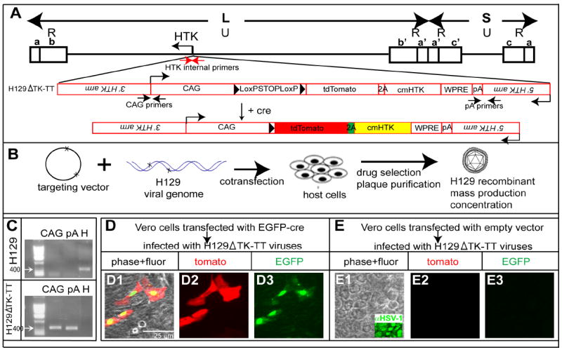
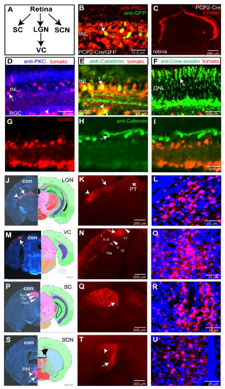
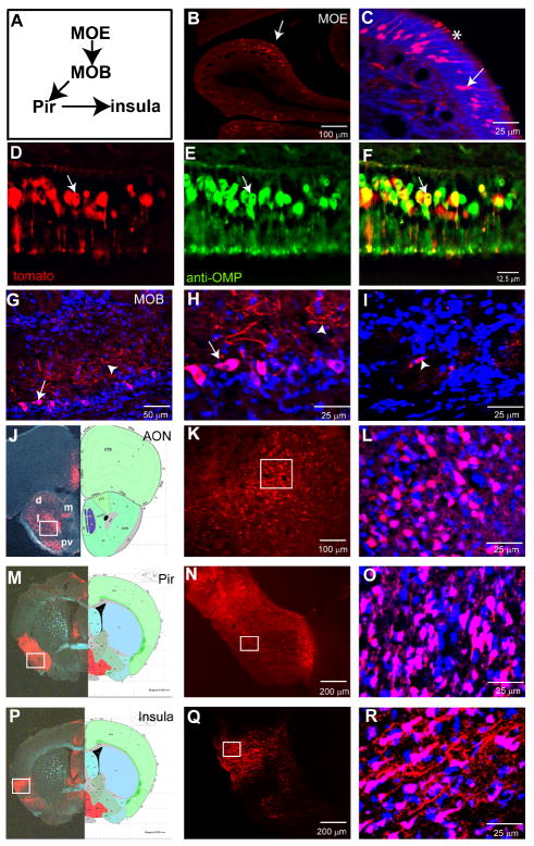
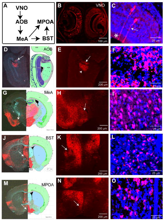
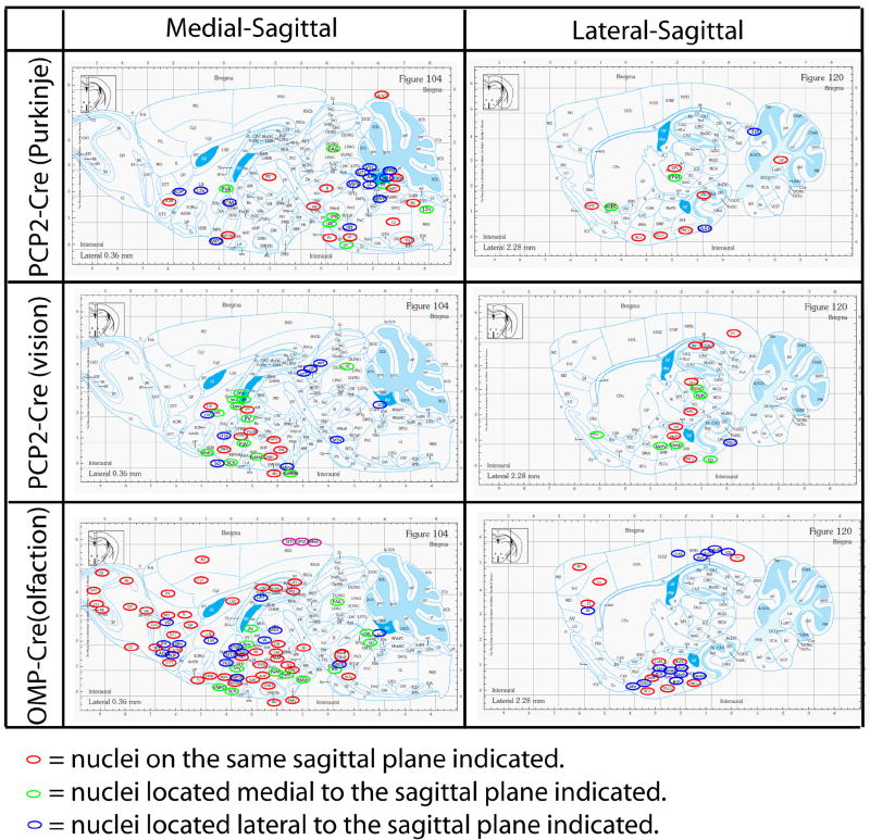
Neurotropic viruses that conditionally infect or replicate in molecularly defined neuronal subpopulations, and then spread transsynaptically, are powerful tools for mapping neural pathways. Genetically targetable retrograde transsynaptic tracer viruses are available to map the inputs to specific neuronal subpopulations, but an analogous tool for mapping synaptic outputs is not yet available. Here we describe a Cre recombinase-dependent, anterograde transneuronal tracer, based on the H129 strain of herpes simplex virus (HSV). Application of this virus to transgenic or knockin mice expressing Cre in peripheral neurons of the olfactory epithelium or the retina reveals widespread, polysynaptic labeling of higher-order neurons in the olfactory and visual systems, respectively. Polysynaptic pathways were also labeled from cerebellar Purkinje cells. In each system, the pattern of labeling was consistent with classical circuit-tracing studies, restricted to neurons, and anterograde specific. These data provide proof-of-principle for a conditional, nondiluting anterograde transsynaptic tracer for mapping synaptic outputs from genetically marked neuronal subpopulations.


Figures

Figure 1
Figure 1. Construction and expression properties of HSV-1 strain H129ΔTK-TT
(A) H129 targeting strategy. Upper, schematic of H129 genome. L, S indicate long and short fragments, respectively; U and R, unique and inverted (a, b, c, and a’, b’ and c’) repeats, respectively. Lower, targeting construct (red). (B) Strategy for generating recombinant H129. (C) Genomic PCR analysis of H129 and H129ΔTK-TT. CAG and pA indicate 3’ and 5’ junction fragments (see (A)). “H” indicates native HTK fragment (see red arrows in A). (D-E) Infection of Vero cells by H129ΔTK-TT with (D) or without (E) EGFP-Cre transfection. Related data regarding the infectivity of H129ΔTK-TT virus is shown in Supplemental Table 2.
Figure 2
Figure 2. H129ΔTK-TT tracing of cerebellar circuits from Purkinje cells
(A) Schematic illustrating the cerebellar-Purkinje pathway. Two-way arrows indicate the olivo-cerebellar tract and cerebello-olivary pathway (Ito, 1984). DCN, deep cerebellar nucleus; VE, vestibular nucleus; RN, red nucleus; IO, inferior olive; VL, ventral lateral thalamic nucleus. (B-C) Intracerebellar injection of wild type (B) or PCP2/L7-Cre transgenic mice (C) with H129ΔTK-TT virus. GFP (C) reports a co-integrated PCP2/L7-EGFP transgene. (D-O) Brain structures labeled with H129ΔTK-TT following recombination in Purkinje cells. DN, dentate nucleus; IP, interposed nucleus; FN, fastigial nucleus; LVe, lateral vestibular nucleus; MVe, medial vestibular nucleus; SuVe, superior vestibular nucleus; V4, 4th ventricle. Left panels in (D, G, J, M) are from injected brains stained for Nissl and native tdT (merged brightfield/fluorescence); right panels are corresponding sections from Allen Reference Atlas (Dong, 2008). Panels (F, I, L, O) are higher magnification images of boxed regions in (E, H, K, N), respectively, from the same or adjacent sections, counter-stained with fluorescent Nissl. Additional labeled areas are shown in Supplemental Figure 1. (P-R) tdTomato labeling is restricted to neurons in the IO. (Q, R), double-labeling for tdT and anti-NeuN (Q) or GFAP (R) antibody staining, in adjacent sections corresponding to the boxed region in (P). Additional data are shown in Supplemental Figure 2 and Supplemental Table 1.
Figure 3
Figure 3. H129ΔTK-TT tracing of visual system circuits from rod bipolar cells
(A) Schematic illustrating visual pathway. (B-I) Retinal sections from PCP2/L7-Cre/GFP mice. (B) GFP and PKCα mark rod bipolar cells (RBCs; arrow) in uninjected mice. (C) Retinal tdT expression following intra-vitreal injection of H129ΔTK-TT virus. (D-I) Note tdT labeling in RBCs (D, arrow), amacrine (E, arrow), and retinal ganglion cells (D, E arrowheads), but not in photoreceptors (F, arrow) or horizontal cells (G-I, arrow). (J-U) Central visual relays labeled by H129ΔTK-TT transport. Left panels in (J, M, P, S) illustrate hemibrains contralateral to injected eye, stained for Nissl and tdT. LGN, lateral geniculate nucleus; PT, pretectal nucleus; VC, visual cortex; (I-VI), layers of visual cortex (N); SC, superior collicullus; SuG, superficial gray of SC; Op, optic nerve layer of SC; DpG; deep gray layer of SC. (S) SCN, suprachiasmatic nucleus; AH, anterior hypothalamus (arrowhead and arrow, respectively, in (T)). (L, O, R, U) are higher magnification views of the labeled areas indicated by arrows in (K, N, Q, T), respectively, counter-stained with fluorescent Nissl. Additional tdT-expressing regions in these mice are shown in Supplemental Figure 3.
Figure 4
Figure 4. H129ΔTK-TT tracing of main olfactory circuits from ORNs
(A) Schematic illustrating main olfactory pathway. MOE, Main Olfactory Epithelium; MOB, Main Olfactory Bulb; Pir, piriform cortex. (B-F) H129ΔTK-TT infected MOE. Arrows (B-C) indicate labeled ORNs, asterisk olfactory cilia. (D-F) section of MOE double-labeled for tdT and anti-OMP; arrows indicate double-positive ORN. (G-I) Labeling in the MOB. (H) higher magnification view of area indicated by arrow in (G). Arrows indicate mitral cells, arrowheads external plexiform layer in (G, H). Arrowhead in (I) indicates periglomerular cell. (J-R) tdT labeling of higher order main olfactory relays. AON, anterior olfactory nucleus (d, m, l, and pv in (J) indicate dorsal, medial, lateral, and posteroventral parts of AON); Pir, piriform cortex; Insula, insular cortex. Organization of panels as in Figure 2D-O. Additional labeled brain areas are shown in Supplemental Figure 4.
Figure 5
Figure 5. H129ΔTK-TT tracing of accessory olfactory circuits
(A) Schematic illustrating VNO system. AOB, accessory olfactory bulb; MeA, medial amygdala; MPOA, medial preoptic area; BST, bed nucleus of the stria terminalis. (B-C) H129ΔTK-TT infected VNO. Arrow (C) indicates labeled olfactory receptor neuron, asterisk olfactory microvilli. (D-O) tdT labeling of higher order accessory olfactory relays. Arrowhead (E) indicates anterior olfactory nuclei. Organization of panels (D-O) as in Fig. 3(J-U).
Figure 6
Figure 6. Comparisons of selected brain structures labeled by H129ΔTK-TT in the visual, olfactory, and cerebellar pathways
Medial-sagittal and lateral-sagittal plates and labeled structures are from the Franklin and Paxinos Atlas (Franklin and Paxinos, 2008). Red circles mark tdT-labeled structures on the indicated saggital planes; green and blue circles mark tdT-labeled structures on adjacent medial and lateral planes, respectively. Related data are shown in Supplemental Figure 5 and Supplemental Table 3.

References

    1. Archin NM, van den Boom L, Perelygina L, Hilliard JM, Atherton SS. Delayed spread and reduction in virus titer after anterior chamber inoculation of a recombinant of HSV-1 expressing IL-16. Invest Ophthalmol Vis Sci. 2003;44:3066–3076. - PubMed
    1. Astic L, Saucier D, Coulon P, Lafay F, Flamand A. The CVS strain of rabies virus as transneuronal tracer in the olfactory system of mice. Brain Res. 1993;619:146–156. - PubMed
    1. Badea TC, Nathans J. Quantitative analysis of neuronal morphologies in the mouse retina visualized by using a genetically directed reporter. J Comp Neurol. 2004;480:331–351. - PubMed
    1. Bak IJ, Markham CH, Cook ML, Stevens JG. Intraaxonal transport of Herpes simplex virus in the rat central nervous system. Brain Res. 1977;136:415–429. - PubMed
    1. Barnett EM, Evans GD, Sun N, Perlman S, Cassell MD. Anterograde tracing of trigeminal afferent pathways from the murine tooth pulp to cortex using herpes simplex virus type 1. J Neurosci. 1995;15:2972–2984. - PMC - PubMed

Publication types

MeSH terms