

Functional specialization of mouse higher visual cortical areas
- PMID: 22196337
- PMCID: PMC3876958
- DOI: 10.1016/j.neuron.2011.11.013
Functional specialization of mouse higher visual cortical areas
Abstract
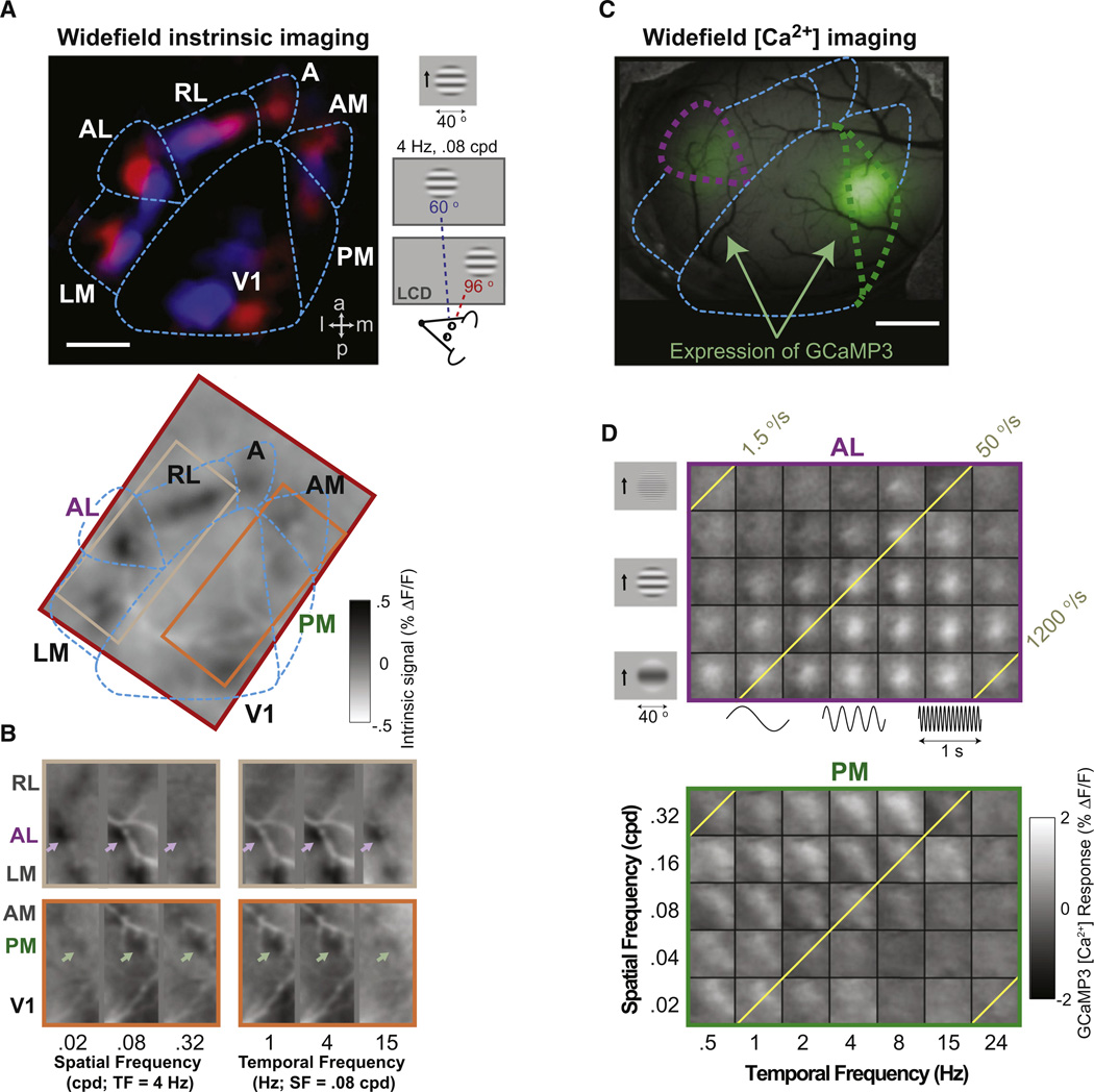
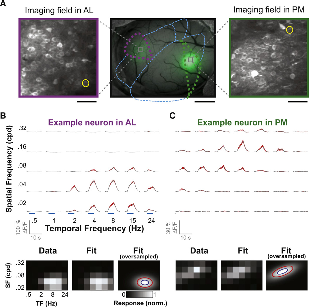
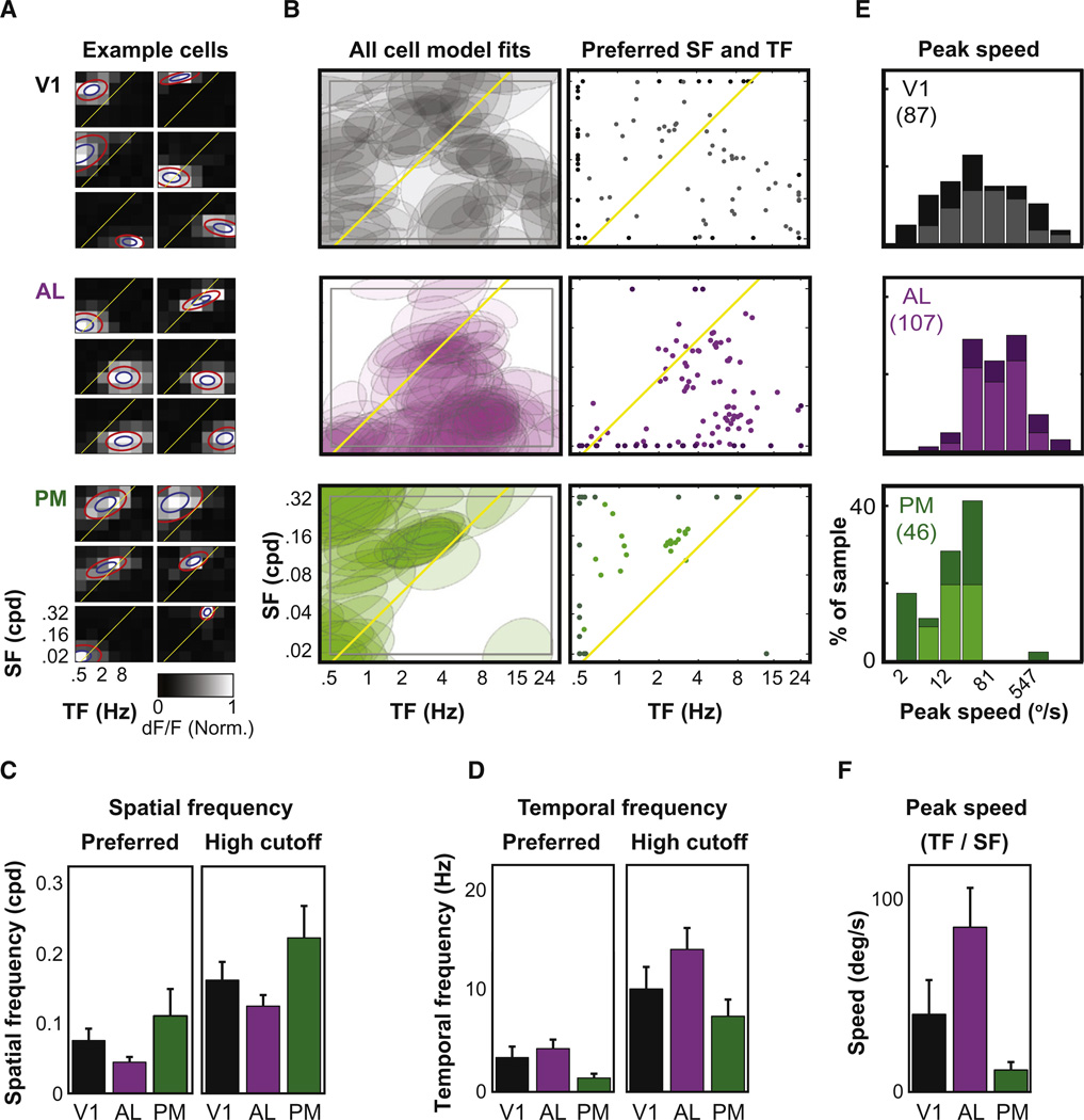
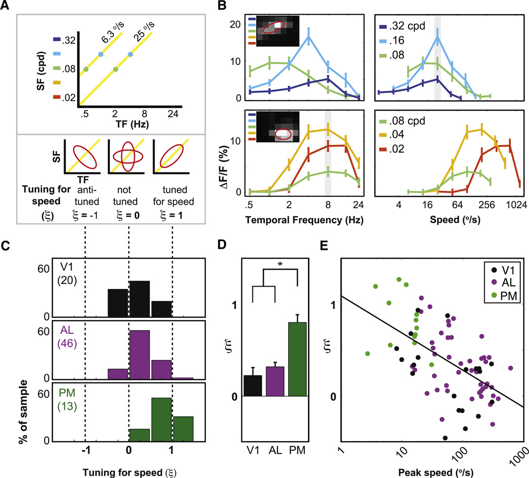
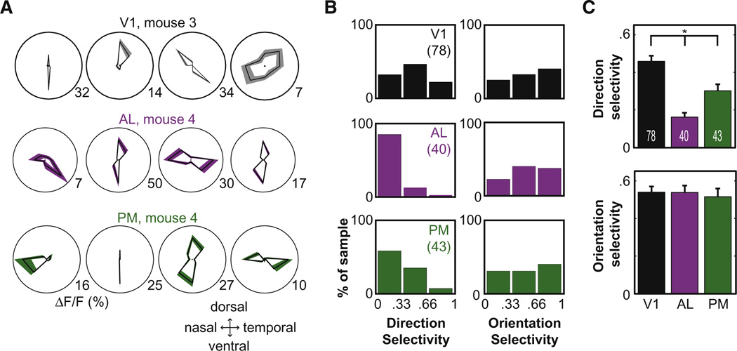
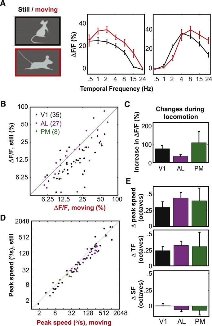
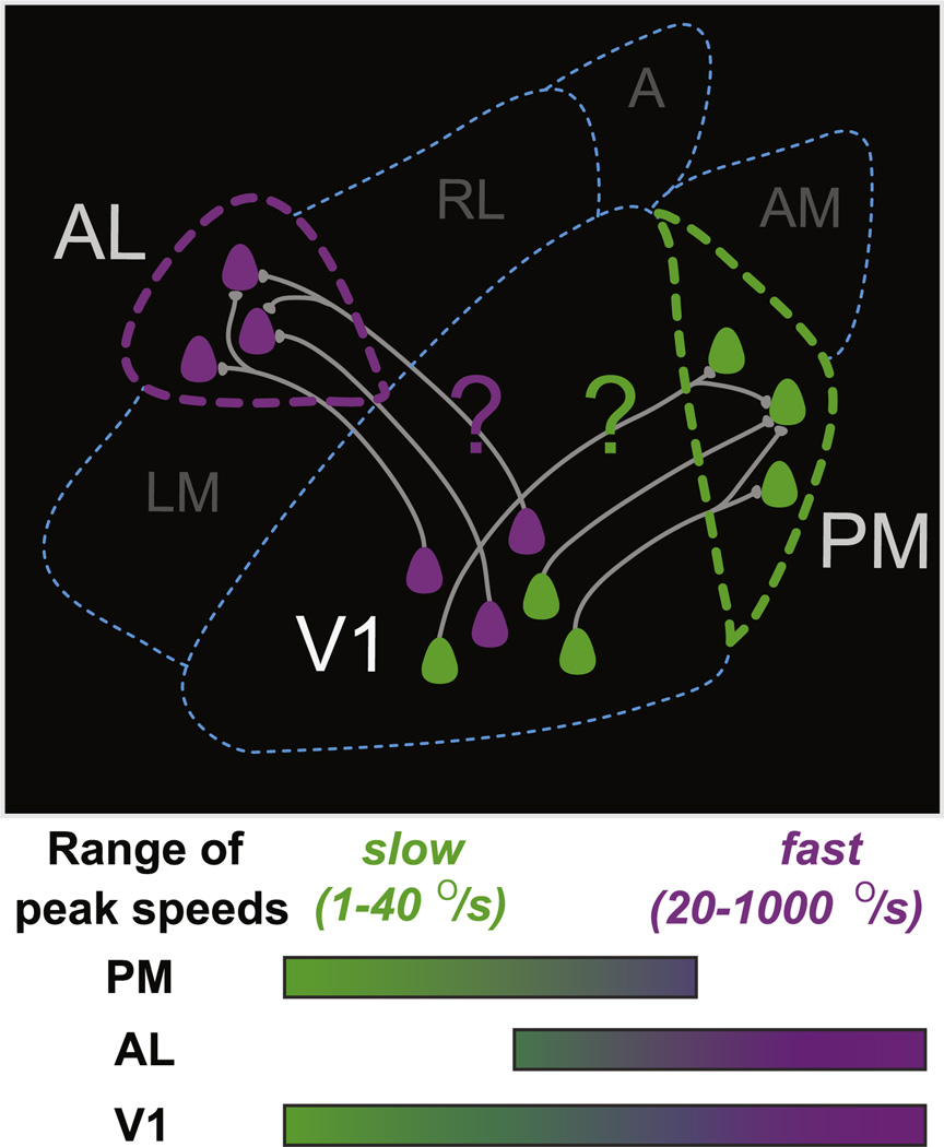
The mouse is emerging as an important model for understanding how sensory neocortex extracts cues to guide behavior, yet little is known about how these cues are processed beyond primary cortical areas. Here, we used two-photon calcium imaging in awake mice to compare visual responses in primary visual cortex (V1) and in two downstream target areas, AL and PM. Neighboring V1 neurons had diverse stimulus preferences spanning five octaves in spatial and temporal frequency. By contrast, AL and PM neurons responded best to distinct ranges of stimulus parameters. Most strikingly, AL neurons preferred fast-moving stimuli while PM neurons preferred slow-moving stimuli. By contrast, neurons in V1, AL, and PM demonstrated similar selectivity for stimulus orientation but not for stimulus direction. Based on these findings, we predict that area AL helps guide behaviors involving fast-moving stimuli (e.g., optic flow), while area PM helps guide behaviors involving slow-moving objects.
Copyright © 2011 Elsevier Inc. All rights reserved.
Figures
Comment in
-
Exploring the next frontier of mouse vision.Neuron. 2011 Dec 22;72(6):889-92. doi: 10.1016/j.neuron.2011.12.011. Neuron. 2011. PMID: 22196324
References
-
- Aggleton JP, Keen S, Warburton EC, Bussey TJ. Extensive cytotoxic lesions involving both the rhinal cortices and area TE impair recognition but spare spatial alternation in the rat. Brain Res. Bull. 1997;43:279–287. - PubMed
-
- Andersen RA, Snyder LH, Bradley DC, Xing J. Multimodal representation of space in the posterior parietal cortex and its use in planning movements. Annu. Rev. Neurosci. 1997;20:303–330. - PubMed
Publication types
MeSH terms
Grants and funding
LinkOut - more resources
Full Text Sources
Other Literature Sources
Molecular Biology Databases
