. 2011 Dec 23;147(7):1511-24.
doi: 10.1016/j.cell.2011.11.032.

A poised chromatin platform for TGF-β access to master regulators

Affiliations

A poised chromatin platform for TGF-β access to master regulators

Qiaoran Xi et al. Cell. .

Abstract

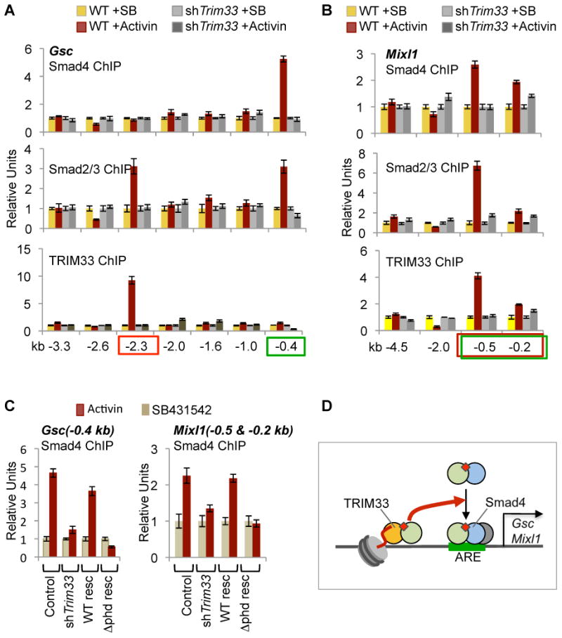
Specific chromatin marks keep master regulators of differentiation silent yet poised for activation by extracellular signals. We report that nodal TGF-β signals use the poised histone mark H3K9me3 to trigger differentiation of mammalian embryonic stem cells. Nodal receptors induce the formation of companion Smad4-Smad2/3 and TRIM33-Smad2/3 complexes. The PHD-Bromo cassette of TRIM33 facilitates binding of TRIM33-Smad2/3 to H3K9me3 and H3K18ac on the promoters of mesendoderm regulators Gsc and Mixl1. The crystal structure of this cassette, bound to histone H3 peptides, illustrates that PHD recognizes K9me3, and Bromo binds an adjacent K18ac. The interaction between TRIM33-Smad2/3 and H3K9me3 displaces the chromatin-compacting factor HP1γ, making nodal response elements accessible to Smad4-Smad2/3 for Pol II recruitment. In turn, Smad4 increases K18 acetylation to augment TRIM33-Smad2/3 binding. Thus, nodal effectors use the H3K9me3 mark as a platform to switch master regulators of stem cell differentiation from the poised to the active state.


Figures

Figure 1
Figure 1. TRIM33 is Engaged in the Nodal Smad Pathway
(A-C) Distribution of TRIM33 in stage E7.5 mouse embryos. Immunohistochemistry analysis of mouse embryo sections with antibodies against TRIM33. Images are (A) whole embryo (30x magnification), (B) node region (150x) and (C) primitive streak (150x) (D) Activin dependent formation of TRIM33-Smad2/3 and Smad4-Smad2/3 complexes in mESCs. mESC cultures were stimulated with activin A (50 ng/ml) for the indicated time periods or treated with SB431542 (SB) (10 μM) to block autocrine nodal like signals. Cell lysates were immunoprecipitated with anti-Smad2/3 antibody and immune complexes were analyzed by immunoblotting using antibodies against the indicated proteins. (E) Formation of the TRIM33-Smad2/3 complex in wild type and Smad4-null mESCs. (F) Formation of Smad4-Smad2/3 complex in wild type and Trim33-depleted mESCs. (G) mESCs that were wild-type, Trim33-depleted, reconstituted with TRIM33, Smad2/3-depleted or Smad4 null were set for EB formation for 2.5 days and then incubated for 2h with activin, SB or no additions. RNA was analyzed by qRT-PCR using primers for the indicated genes. Data are the mean ± S.D of quadruplicates and represent three independent experiments. (H) Wild type or Trim33-null mESCs were treated and analyzed as described in (G). (I) Schematic representation of TRIM33 and Smad4 dependent nodal/activin gene responses. (J) Control, Trim33-depleted and Smad4-null ESCs were set for EB formation for 2.5 days and processed as in (G). Total RNA was subjected to genome-wide transcriptiomic analysis with MOE 430A 2.0 microarray. The Venn diagram shows a summary of TRIM33 and Smad4-dependent and -independent activin responsive genes (>3 fold change, p<0.05). For additional information related to how TRIM33 participates in activin and TGF-β responses, see Figure S1.
Figure 2
Figure 2. TRIM33 is Required for Mesendodermal Differentiation of Mammalian ESCs
(A) Schematic summary of mouse ESCs differentiation in culture (Murry and Keller, 2008). (B) Trim33-null mESCs were verified by immunoblot analysis using antibody against TRIM33. (C-F) Expression pattern of three germ layer markers in mESC differentiation. Wild type and Trim33-null mESCs (two clones each) were set for EB formation for the indicated lengths of time. Total RNA was analyzed by qRT-PCR using primers for the indicated genes. Data are the mean ± S.D of quadruplicates and are representative of two independent experiments. (G) TRIM33 was depleted in human ESCs using lentiviral shRNA vector (refer to Figure S2I). Control and TRIM33-knockdown were set for mesoderm differentiation and total RNA was analyzed by qRT-PCR using primers for the indicated genes. Data are the mean ± S.D of quadruplicates and are representative of two independent experiments. (H) Immunofluorescence analysis of mesendoderm markers Foxa2 and T/Brachyury, and ectoderm marker Nestin in wild-type and Trim33-depleted mouse EBs at the indicated times. For additional information related to the role of TRIM33 in ESC differentiation, see Figure S2.
Figure 3
Figure 3. TRIM33 Recognizes a Dual Histone Mark Typical of Poised Chromatin
(A) Representation of TRIM33 protein domains (top panel) and summary of H3 histone binding specificity of the PHD-Bromo cassette based on the present results (lower panel). (B-C) Requirement of the PHD finger of TRIM33 for induction of Gsc and Mixl1 by activin (B) or by nodal-like signals in the media (C). mESCs that were wild type, Trim33-depleted or reconstituted with the indicated TRIM33 constructs, were set for EB formation for 2.5 days and then incubated with activin or SB431542 for 2h. Total RNA was analyzed by qRT-PCR using primers for the indicated genes. Data are the mean ± S.D of quadruplicates and are representative of three independent experiments. (D-E, and G) Binding assays with biotinylated H3 histone peptides of the indicated lengths and modifications. Peptides were coupled to avidin-agarose and used as bait to pull down Flag-tagged TRIM33 generated in HEK293T cells (D, G) or a bacterially expressed, GST-tagged C-terminal fragment of TRIM33 (E). Bound TRIM33 was detected by immunoblotting. (F) ITC-based measurements indicate that TRIM33 PHD finger prefers to bind histone H3(1-15) containing unmodified lysine 4 and trimethylated lysine 9. Dissociation constants are listed. (H) ITC-based measurements of binding affinities of the TRIM33 PHD-Bromo cassette for H3(1-28) peptides containing combinations of modifications. Dissociation constants are listed. For additional information related to how TRIM33 recognizes poised chromatin, see Figure S3.
Figure 4
Figure 4. Structural Basis for TRIM33 PHD-Bromo Cassette Binding to Histone H3 Marks
(A) Superposition of the crystal structures of the PHD-Bromo cassettes of TRIM24 (cyan; PDB code: 3O33; 2.0 Å) and TRIM33 (magenta; this study; 3.0 Å) in the free state. There is an additional α-helical segment extending from αB of Bromo in TRIM33 (dashed circle) relative to TRIM24. (B) The 2.7 Å crystal structure of H3(1-22)K9me3-K14acK18ac peptide bound to a single TRIM33 PHD-Bromo cassette. The peptide is shown in a yellow stick representation, together with labeled N-terminus, and side chains of unmethylated K4, K9me3 and K18ac. The TRIM33 PHD-Bromo is shown in a ribbon representation, with PHD in cyan and Bromo in brown colors. (C) Detailed interactions between 1-9 segment of H3(1-22)K9me3-K14acK18ac peptide (in yellow) and the PHD finger (in cyan) of the TRIM33 PHD-Bromo cassette in the complex. The side chains of residues 1 to 9 of the peptide are shown and labeled, together with PHD finger backbone and side chain residues involved in intermolecular hydrogen bond formation (dashed red lines). The two zinc ions of the PHD finger are indicated by silver balls. (D) Recognition of R17-K18ac segment of H3(1-22)K9me3/K14ac/K18ac peptide (in yellow) by Bromo (in brown) of the TRIM33 PHD-Bromo cassette in the complex. A hydrogen bond between Arg17 of the peptide and Glu981 of Bromo is shown by a dashed red line. (E) ITC-based measurements of binding affinities of three mutant-forms of TRIM33 PHD-Bromo and their association with H3(1-28)K9me3K18ac histone peptide. For additional structural details of acetyllysine and K9me3 binding pockets, see Figure S4.
Figure 5
Figure 5. Signal-driven Binding of TRIM33 to H3K9me3 Domains of theGsc and Mixl1 Promoters
(A) Scheme of the Gsc region spanning 3.3kb upstream of the transcription start site, and sequence conservation plot between the human and mouse genomes (VISTA graphs: conserved non-coding sequence is shown in pink). The positions of the ARE (Labbé et al., 1998) non-ARE SBEs, and H3K9me3-rich region (present results) are shown. (B) ChIP analysis of mESCEBs at day 2.5 using antibodies against the indicated histone H3 modified residues in the Gsc promoter region. Agarose protein A (for rabbit antibodies) or G (for mouse antibodies) beads alone were used as negative controls. Data in (B-D and F) were obtained by qRT-PCR of regions of interest in Gsc or Mixl1, and data are the mean ± S.D. of quadruplicates and represent at least three independent experiments. (C) ChIP analysis of Gsc promoter regions was performed as indicated in (B) using mono-nuleosome preparations as template. (D) Wild type or Smad2/3-depleted mESC EBs at day 2.5 were incubated with activin or SB 431542 for 2 h, ChIP analysis of the Gsc promoter region was then performed using anti-TRIM33 and Smad2/3 antibodies. (E) Scheme of the Mixl1 region spanning 4.5 kb upstream of the transcription start site, and sequence conservation plot between the human and mouse genomes (VISTA graphs: conserved non-coding sequence is shown in pink). The positions of the ARE (Hart et al., 2005) non-ARE SBEs, and H3K9me3-rich region (present results) are shown. (F) mESC EBs at day 2.5 were incubated for 2 h with the indicated additions. ChIP analysis of the Mixl1 promoter region was performed using antibodies against the indicated proteins. (G) Schematic summary of signal-induced TRIM33-Smad2/3 and Smad4-Smad2/3 complexes binding to H3k9me3-K18ac and ARE sites, respectively, in the Gsc and Mixl1 promoters. For additional information related to the Mixl1 promoter, see Figure S5.
Figure 6
Figure 6. Smad2/3-Smad4 Binding to AREs in Poised Promoters Required TRIM33
(A-B) Wild type or Trim33-depleted mESCs were set to form EBs for 2.5 days, and were then incubated for 2 h with the indicated additions. ChIP analysis of the Gsc promoter (A) or Mixl1 promoter (B) was performed using antibodies against the indicated proteins. Data are the mean ± S.D. of quadruplicates and are representative of at least three independent experiments. (C) mESCs that were wild type, Trim33-depleted, or reconstitutes with wild-type or truncated TRIM33 constructs were set to form EBs for 2.5 days. ChIP analysis of the Gsc(-0.4kb) and Mixl1(combined -0.5 kb and -0.2 kb)promoters was done using antibodies against Smad4. Data are the mean ± S.D. of quadruplicates and represent three independent experiments. (D) Schematic summary: signal-driven binding of TRIM33 and Smad2/3 to H3K9me3-rich regions of Gsc and Mixl1 enables binding of Smad4 and Smad2/3 to the AREs.
Figure 7
Figure 7. TRIM33 and SMAD Cooperation in Activation of Master Regulators
(A) mESCs that were wild type, Smad2/3-depleted, or Smad4-null were set to form EBs for 2.5 days, and then treated for 2h with activin or SB431542. ChIP analysis of the Gsc promoter was done using antibodies against TRIM33. Data are the mean ± S.D. of quadruplicates and representative of three independent experiments. (B) mESCs that were wild type or Smad4-null were treated as in (A), and ChIP analysis of the Gsc and Mixl1 promoters was done using antibodies against H3K18ac, Data are the mean ± S.D. of quadruplicates and are representative of two independent experiments. (C) TRIM33 competes with HP1γ for binding to H3K9me-K18ac. Biotinylated histone H3 peptides with the indicated modifications were used in pull-down binding assays with a fixed amount of Flag-tagged HP1γ protein and an increasing amount of Flag-tagged TRIM33. Bead-bound proteins were analyzed by immunoblotting. (D) Activin-induced displacement of HP1γ from TRIM33 binding sites in the Gsc and Mixl1 promoters, but not in Trim33-null EBs. mESC wild type and Trim33-null EBs at day 2.5 were incubated with activin or SB for 2 h. ChIP assays were done using antibodies against HP1γ (brown bars) or no antibody (blue bars), and qRT-PCR primers amplifying the −2.3 kb region of Gsc or the −0.2 and -0.5kb kb regions of Mixl1. Data are the mean ± S.D. of quadruplicates and are representative of two independent experiments. (E) Activin increases RNA Pol II binding to the Gsc and Mixl1 promoters, but not in Trim33-null EBs. The EB samples used in (D) were subjected to ChIP using antibodies against RNA Pol II and qRT-PCR primers amplifying the -0.1 kb region of Gsc or the -0.2 kb region of Mixl1. Data are the mean ± S.D. of quadruplicates and are representative of two independent experiments. (F) Schematic summary of signal-driven Smad activation of master regulators in embryonic stem cells. Gsc and Mixl1 present key features of the poised state in their promoters, Including the H3K9me3 mark of quiescent chromatin and the chromatin compacting factor HP1γ bound to this region. Two nodal-induced Smad complexes cooperate to promote switching of the master regulators Gsc and Mixl1 from poised to activated state in response to nodal-like signals, thereby committing embryo cells to mesendodermal fate. See Discussion for more details. For additional information related to the Smad4-dependent enhancement of TRIM33 binding, see Figure S6.

References

    1. Agricola E, Randall RA, Gaarenstroom T, Dupont S, Hill CS. Recruitment of TIF1gamma to Chromatin via Its PHD Finger-Bromodomain Activates Its Ubiquitin Ligase and Transcriptional Repressor Activities. Mol Cell. 2011;43:85–96. - PubMed
    1. Alarcón C, Zaromytidou AI, Xi Q, Gao S, Yu J, Fujisawa S, Barlas A, Miller AN, Manova-Todorova K, Macias MJ, et al. Nuclear CDKs drive Smad transcriptional activation and turnover in BMP and TGF-beta pathways. Cell. 2009;139:757–769. - PMC - PubMed
    1. Aragón E, Goerner N, Zaromytidou AI, Xi Q, Escobedo A, Massagué J, Macias MJ. A Smad action turnover switch operated by WW domain readers of a phosphoserine code. Genes Dev. 2011;25:1275–1288. - PMC - PubMed
    1. Bai X, Kim J, Yang Z, Jurynec MJ, Akie TE, Lee J, LeBlanc J, Sessa A, Jiang H, DiBiase A, et al. TIF1gamma controls erythroid cell fate by regulating transcription elongation. Cell. 2010;142:133–143. - PMC - PubMed
    1. Bardeesy N, Cheng KH, Berger JH, Chu GC, Pahler J, Olson P, Hezel AF, Horner J, Lauwers GY, Hanahan D, et al. Smad4 is dispensable for normal pancreas development yet critical in progression and tumor biology of pancreas cancer. Genes Dev. 2006;20:3130–3146. - PMC - PubMed

Publication types

Associated data