. 2011 Dec 23;147(7):1615-27.
doi: 10.1016/j.cell.2011.11.027.

The functional organization of cutaneous low-threshold mechanosensory neurons

Affiliations

The functional organization of cutaneous low-threshold mechanosensory neurons

Lishi Li et al. Cell. .

Abstract

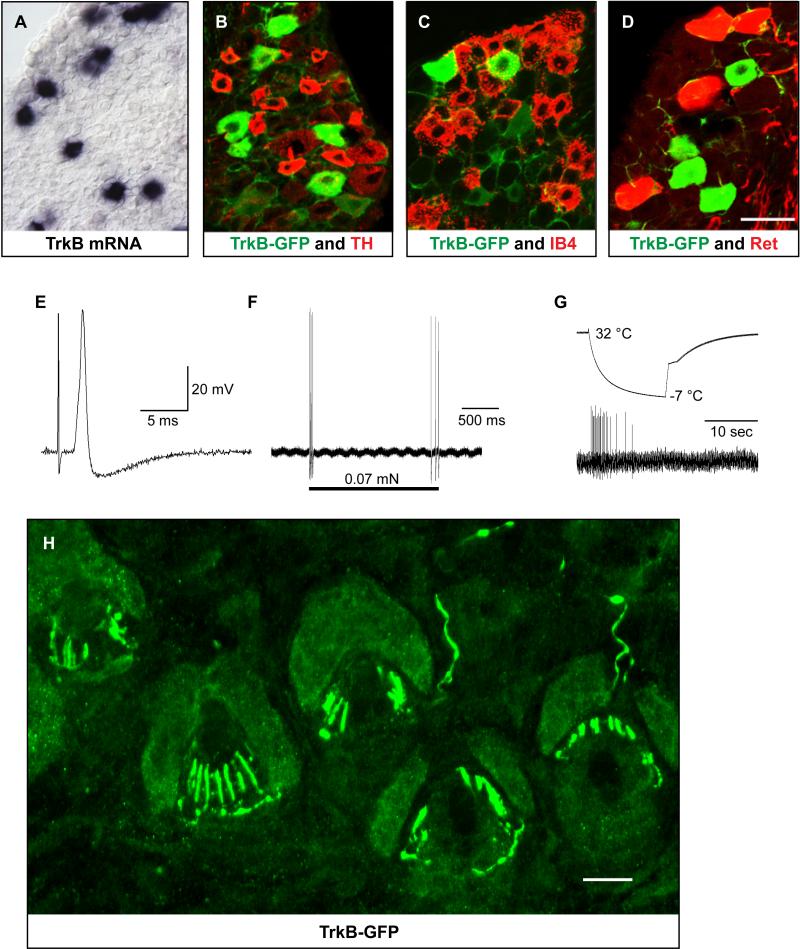
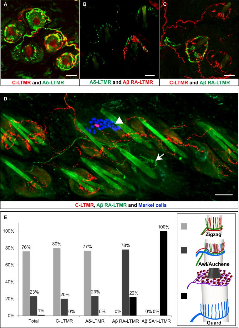
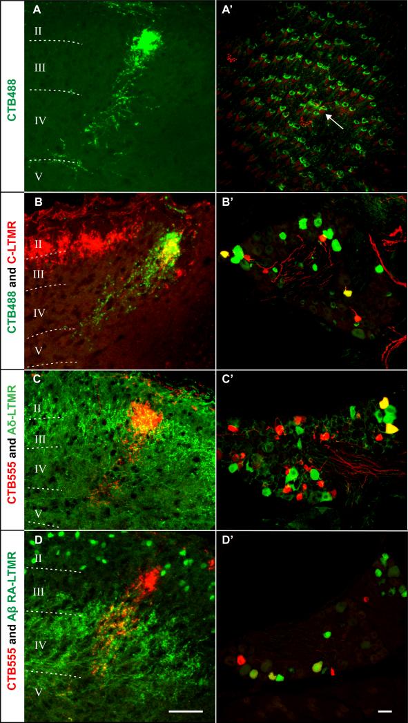
Innocuous touch of the skin is detected by distinct populations of neurons, the low-threshold mechanoreceptors (LTMRs), which are classified as Aβ-, Aδ-, and C-LTMRs. Here, we report genetic labeling of LTMR subtypes and visualization of their relative patterns of axonal endings in hairy skin and the spinal cord. We found that each of the three major hair follicle types of trunk hairy skin (guard, awl/auchene, and zigzag hairs) is innervated by a unique and invariant combination of LTMRs; thus, each hair follicle type is a functionally distinct mechanosensory end organ. Moreover, the central projections of Aβ-, Aδ-, and C-LTMRs that innervate the same or adjacent hair follicles form narrow LTMR columns in the dorsal horn. These findings support a model of mechanosensation in which the activities of Aβ-, Aδ-, and C-LTMRs are integrated within dorsal horn LTMR columns and processed into outputs that underlie the perception of myriad touch sensations.


Figures

Figure 1
Figure 1. The TH+ adult DRG neurons are C-LTMRs and form longitudinal lanceolate endings associated with zigzag and awl/auchene hair follicles
(A-D) Double immunostaining of TH and NFH (A) (n=8), TH and CGRP (B) (n=6), TH and cRet (C) (n=4), and TH and IB4 (D) (n=7) on adult thoracic DRG sections. TH+ DRG neurons do not express NFH (<1%) or CGRP (<5%). The majority of TH+ neurons express cRet (>90%) but do not bind to the lectin IB4 (<1%). (E-F) Intracellular recordings of TH+ DRG neurons were performed using the ex vivo skin nerve preparation. Small-diameter DRG neurons were randomly selected and recorded (n=21). The recorded neurons were then labeled with Neurobiotin, and co-stained with TH and GFRα2 or TH and IB4. Among the recorded neurons, all of those that exhibited classically defined features of C-LTMRs (n=5; See text) (E) are TH+, GFRα2+ and IB4- (F) (arrows). (G) THCreER; Rosa26IAP mice were treated with 4-HT (1mg per day for three days, from P13 to P15, by oral gavage). Double immunostaining of AP and TH shows that a small number of TH+ DRG neurons (~10%) were labeled with AP (n=2). (H-K) Whole-mount AP staining of thoracic DRGs and trunk hairy skin shows that DRG cell bodies (H) and their peripheral endings in the skin (I) are sparsely labeled. Individual axonal branches arborize and form longitudinal lanceolate endings (J and K) (n=26). (L) The longitudinal lanceolate endings of the C-LTMRs are similarly labeled in THCreER;Rosa26LSLtdTomato mice, which enables double labeling of different LTMR endings (n=24). Whole-mount immunostaining of the dsRed signal of hairy skin showed that 80% of C-LTMR lanceolate endings are associated with zigzag hair follicles and 20% with awl/auchene hair follicles, while none were observed to associate with guard hair follicles (n=212 hair follicles examined). Scale bars: 50 μm (A-D) and (G and H); 100 μm (I and J); 20 μm (F) and (K and L).
Figure 2
Figure 2. The TrkB+ adult DRG neurons are Aδ-LTMRs and form longitudinal lanceolate endings associated with zigzag and awl/auchene hair follicles
(A) In situ hybridization for TrkB mRNA using adult thoracic DRG sections shows that TrkB is expressed in a subset of medium-diameter DRG neurons. (B-D) Double staining of GFP and TH (B) or GFP and IB4 (C) on thoracic DRG sections from TrkBtauEGFP mice shows that TrkB+ DRG neurons are distinct from TH+ C-LTMRs and IB4+ nonpeptidergic nociceptors. Double immunostaining of GFP and dsRed on thoracic DRG sections from RetCreERT2; Rosa26LSL-tdTomato; TrkBtauEGFP mice (D) shows that TrkB+ neurons are distinct from early Ret+ Aβ RA-LTMRs. (E-G) The ex vivo skin-nerve preparation was used to assess mechanically evoked responses and physiological properties of individual TrkBtauEGFP+ neurons. The TrkBtauEGFP+ neurons exhibit exquisite sensitivity and rapid adaptation to mechanical stimulation (von Frey threshold < 0.07 mN) (E and F) and an average conduction velocity of 5.8 ± 0.9 m/s (n = 19). These neurons also responded to cooling but not warming of the skin (G). The average receptive field size of these neurons is 2 × 2 mm2. Nineteen neurons from 7 different animals were recorded and each displayed physiological properties that are characteristic of D-hair cells, or Aδ-LTMRs. (H) Anti-GFP immunostaining of a section of back hairy skin from TrkBtauEGFP mice shows that the GFP+ neurons form longitudinal lanceolate endings (n=20). 77% of the GFP+ lanceolate endings associate with zigzag hair follicles while the remaining 23% associate with awl/auchene hair follicles (n=50 hair follicles examined). Scale bars: 20 μm.
Figure 3
Figure 3. The Aβ RA-LTMRs are labeled with GFP in Npy2r-GFP mice and their GFP+ peripheral axons form longitudinal lanceolate endings associated with guard and awl/auchene hairs
(A-B) In Npy2r-GFP mice, double immunostaining of GFP and NFH shows that the GFP+ neurons have large-diameter somata and express NFH (n=7). Double staining of GFP and TH shows that Npy2r-GFP+ neurons are distinct from the C-LTMRs (n=5). (C) In Npy2r-tdTomato; TrkBtauEGFP mice, double immunostaining of dsRed and GFP shows that the Npy2r-GFP+ neurons are distinct from the Aδ-LTMRs (n=5). (D and E) The ex vivo skin-nerve preparation was used to assess mechanically evoked responses and the physiological properties of individual Npy2r-GFP+ neurons. Intracellular recordings were performed in the same manner as those described for TrkBtauEGFP animals. 18 neurons from 5 different animals were recorded and exhibited low mechanical thresholds (0.07 mN), rapid conduction velocities (13.6 ± 2.7 m/sec) and rapidly adapting responses, which are the hallmark features of Aβ RA-LTMRs. (F and G) In hairy skin sections from Npy2r-GFP mice, double immunostaining of GFP and MBP (F) (n=2) and GFP and S100 (G) (n=4) shows that Npy2r-GFP+ neurons form longitudinal lanceolate endings associated with hair follicles. These GFP+ endings have MBP+ myelinated parental axons and unmyelinated terminals that are intimately associated with S100+ terminal Schwann cells (G). (H and I) Whole-mount immunostaining of GFP and the Merkel cell marker Troma1 of hairy skin from Npy2r-GFP mice showed that 22% of the GFP+ lanceolate endings are associated with guard hair follicles, as determined by the presence of Troma1+ Merkel cells at the follicle mouth (H) (n=6); 78% of the GFP+ lanceolate endings are associated with awl/auchene hair follicles, which are smaller in size and do not have Merkel cells (I); zigzag hair follicles are devoid of GFP+ endings (n=128 hair follicles examined). Scale bars: 20 μm (A-C, F-I).
Figure 4
Figure 4. The organization of peripheral axonal endings of Aβ-LTMRs, Aδ-LTMRs, and C-LTMRs in hairy skin
(A) In hairy skin sections from THCreER ; Rosa26LSL-tdTomato ; TrkBtauEGFP mice, TH+ C-LTMR axonal endings are labeled with tdTomato fluorescence (red) and TrkB+ Aδ-LTMR axonal endings are labeled by GFP immunostaining (green). Virtually every zigzag and awl/auchene hair receives interdigitated C-LTMR and Aδ-LTMR longitudinal lanceolate endings (n=3). (B) In hairy skin sections from Npy2r-tdTomato; TrkBtauEGFP mice, Aβ RA-LTMR and TrkB+ Aδ-LTMR axonal endings are labeled by dsRed (red) and GFP (green) immunostaining, respectively. Awl/auchene hairs exhibit interdigitated Aδ-LTMR and Aβ RA-LTMR longitudinal lanceolate endings whereas zigzag hairs have Aδ-LTMR endings but not Aβ RA-LTMR endings (n=5). (C) In hairy skin sections from THCreER ; Rosa26LSL-tdTomato ; Npy2r-GFP mice, TH+ C-LTMR axonal endings are labeled with tdTomato fluorescence (red) and Aβ RA-LTMR axonal endings are labeled by GFP immunostaining (green). Awl/auchene hairs exhibit interdigitated C-LTMR and Aβ RA-LTMR longitudinal lanceolate endings whereas zigzag hairs have C-LTMR endings but not Aβ RA-LTMR endings (n=5). (D) Whole-mount immunostaining of dsRed, GFP, and Troma1 of hairy skin from THCreER ; Rosa26LSL-tdTomato ; Npy2r-GFP mice reveals the organization of C-LTMR and Aβ RA-LTMR axonal endings around guard hair follicles (n=3). Shown is a representative guard hair follicle associated with a cluster of Troma1+ Merkel cells at the mouth (blue, arrowhead) and innervated by GFP+ Aβ RA-LTMR longitudinal lanceolate endings (green, arrow). Surrounding the guard hair follicle are zigzag and awl/auchene hair follicles, which are innervated by C-LTMRs (red). (E) Left: quantification of the percentages of zigzag (light grey bars), awl/auchene (dark grey bars) or guard hairs (black bars) among total hairs (n=1493), hairs innervated by C-LTMRs (n=212), hairs innervated by Aδ-LTMRs (n=50), hairs innervated by Aβ RA-LTMRs (n=128), and hairs innervated by Aβ, SA1-LTMRs (n=15). Right: schematic pictures of zigzag, awl/auchene and guard hair follicles showing their unique combinations of LTMR endings. Scale bars: 20 μm (A-C); 50 μm (D).
Figure 5
Figure 5. The central projections of C-, Aδ-, and Aβ RA-LTMRs terminate in distinct, but partially overlapping laminae of the spinal cord dorsal horn
(A) In THCreER; Rosa26LSL-tdTomato mice, tdTomato fluorescence shows axonal terminals of individual C-LTMRs located within lamina IIiv of the spinal cord dorsal horn (20-30 μm mediolaterally, 30-40 μm dorsoventrally) (n=24). (B) In TrkBtauEGFP mice, GFP fluorescence shows central projections of TrkB+ Aδ-LTMRs mainly restricted to lamina III (50-60 μm dorsoventrally) (n=9). (C) In Npy2r-GFP mice, GFP immunostaining shows central endings of Aβ RA-LTMRs located in lamina III through V (~100 μm dorsoventrally) (n=8). (D) In spinal cord sections from THCreER; Rosa26LSL-tdTomato ; TrkBtauEGFP mice, the central terminals of C-LTMRs labeled by tdTomato fluorescence (red) are located dorsally, but partially overlapping with the central projections of Aδ-LTMRs, which are visualized by GFP fluorescence (green) (n=3). (E) In spinal cord sections from THCreER ; Rosa26LSL-tdTomato ; Npy2r-GFP mice, TH+ C-LTMR axonal endings labeled with tdTomato fluorescence (red) are located dorsal to the central projections of Aβ RA-LTMRs labeled by GFP immunostaining (green) (n=5). (F) In spinal cord sections from RetCreERT2;RosaLSLtdTomato ; TrkBtauEGFP mice, the central projections of Aδ-LTMRs labeled by GFP fluorescence (green) are located dorsally, but partially overlapping with the central projections of Aβ RA-LTMRs, which are labeled by dsRed immunostaining (red) (n=3). Similar findings were made in experiments that used Npy2r-tdTomato; TrkBtauEGFP mice (data not shown). (G) A schematic of a transverse section of thoracic spinal cord showing the relative positions of the central projections of C-LTMRs, Aδ-LTMRs, and Aβ RA-LTMRs in the dorsal horn. Scale bars: 50 μm (A-F)
Figure 6
Figure 6. LTMR columns of the spinal cord dorsal horn
(A-A’) The central (spinal cord dorsal horn) terminations of sensory neurons whose peripheral axonal endings are labeled by microinjection of CTB488 into a small region of back hairy skin (n=80). The size of the dorsal horn axonal termination region for CTB488-labeled neurons is 20-30 μm along the mediolateral axis and ~200 μm along the dorsoventral axis (A). In the example shown (A’), a 1.02 mm2 area of skin containing 123 hair follicles was labeled with CTB488 (green) followed with whole-mount immunostaining of Troma1 (red) (n=9). A single guard hair (arrow) that associates with Troma1+ Merkel cells is located in the middle of the injection area. On average, 190 ± 23 hair follicles were labeled by microinjection of CTB488 into a small region of back hairy skin (n=11). (B-D) CTB488 or CTB555 were injected into a small region of back hairy skin of mice in which the different populations of LTMRs are genetically labeled. CTB labeled central afferents overlap with C-LTMR central terminals in THCreER; Rosa26LSL-tdTomato mice (B) (n=4), Aδ-LTMR central projections in TrkBtauEGFP mice (C) (n=5) and Aβ RA-LTMR central projections in Npy2r-GFP mice (D) (n=4). (B’-D’) A small number of cell bodies of each LTMR type are retrogradely labeled by CTB microinjection into the skin. CTB488 (green) and tdTomato fluorescence (red) in a thoracic DRG section from a THCreER; Rosa26LSL-tdTomato mouse (B’) (n=4). CTB555 (red) and GFP fluorescence (green) in a thoracic DRG section of a TrkBtauEGFP mouse (C’) (n=5). CTB555 (red) and GFP fluorescence (green) in a thoracic DRG sections from an Npy2r-GFP mouse (D’) (n=3). Scale bars: 200 μm (A’), 50 μm (A-D) and (B’-D’).
Figure 7
Figure 7. The organization of LTMR endings in hairy skin and the spinal cord dorsal horn
The peripheral endings of Aβ-LTMRs, Aδ-LTMRs and C-LTMRs associate with either one or two of the three types of hair follicles of trunk and proximal limb hairy skin. At zigzag hair follicles, C-LTMRs (red) and Aδ-LTMRs (green) form interdigitated longitudinal lanceolate endings; At awl/auchene hair follicles, Aβ RA-LTMRs (blue), Aδ-LTMRs (green), and C-LTMRs (red) form interdigitated longitudinal lanceolate endings; Guard hair follicles are associated with longitudinal lanceolate endings formed by Aβ RA-LTMRs (blue) and clusters of Merkel cells, or touch domes and thus Aβ SA1-LTMRs (purple). The central terminals of LTMRs that innervate the same or adjacent hair follicles within a peripheral LTMR unit are aligned to form a narrow LTMR column in the spinal cord dorsal horn.

References

    1. Badea TC, Hua ZL, Smallwood PM, Williams J, Rotolo T, Ye X, Nathans J. New mouse lines for the analysis of neuronal morphology using CreER(T)/loxP-directed sparse labeling. PloS One. 2009;4:e7859. - PMC - PubMed
    1. Bessou P, Burgess PR, Perl ER, Taylor CB. Dynamic properties of mechanoreceptors with unmyelinated (C) fibers. J Neurophysiol. 1971;34:116–131. - PubMed
    1. Brown AG. Organization in the spinal cord: the anatomy and physiology of identified neurones. illustrated edn. Springer-Verlag; Berlin ; New York: 1981a. 1981.
    1. Brown AG. The spinocervical tract. Prog Neurobiol. 1981b;17:59–96. - PubMed
    1. Brown AG, Franz DN. Responses of spinocervical tract neurones to natural stimulation of identified cutaneous receptors. Exp Brain Res. 1969;7:231–249. - PubMed

Publication types