

Reprogramming cardiomyocyte mechanosensing by crosstalk between integrins and hyaluronic acid receptors
- PMID: 22196970
- PMCID: PMC3386849
- DOI: 10.1016/j.jbiomech.2011.11.023
Reprogramming cardiomyocyte mechanosensing by crosstalk between integrins and hyaluronic acid receptors
Abstract
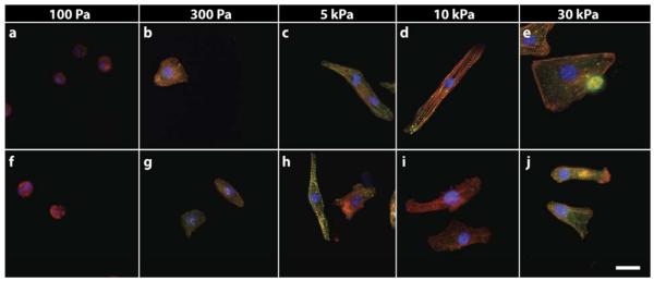
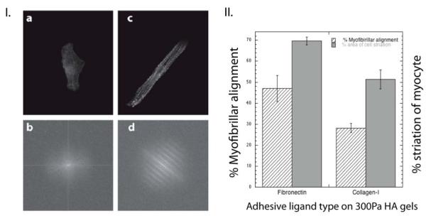
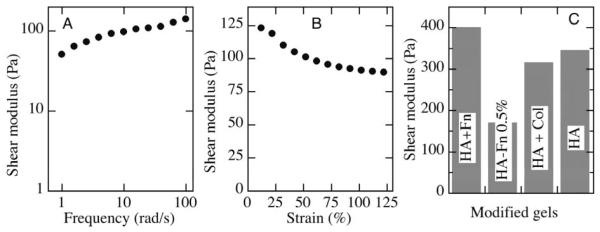
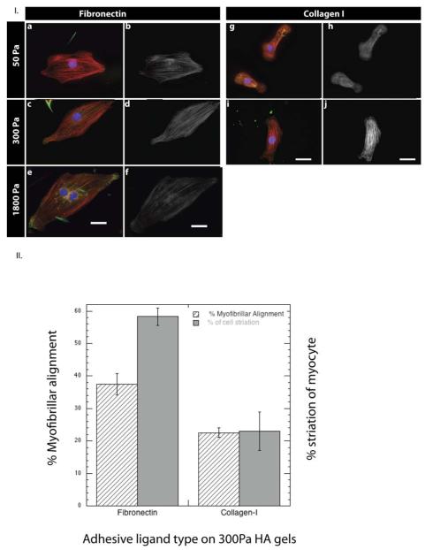
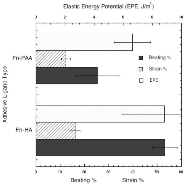
The elastic modulus of bioengineered materials has a strong influence on the phenotype of many cells including cardiomyocytes. On polyacrylamide (PAA) gels that are laminated with ligands for integrins, cardiac myocytes develop well organized sarcomeres only when cultured on substrates with elastic moduli in the range 10 kPa-30 kPa, near those of the healthy tissue. On stiffer substrates (>60 kPa) approximating the damaged heart, myocytes form stress fiber-like filament bundles but lack organized sarcomeres or an elongated shape. On soft (<1 kPa) PAA gels myocytes exhibit disorganized actin networks and sarcomeres. However, when the polyacrylamide matrix is replaced by hyaluronic acid (HA) as the gel network to which integrin ligands are attached, robust development of functional neonatal rat ventricular myocytes occurs on gels with elastic moduli of 200 Pa, a stiffness far below that of the neonatal heart and on which myocytes would be amorphous and dysfunctional when cultured on polyacrylamide-based gels. The HA matrix by itself is not adhesive for myocytes, and the myocyte phenotype depends on the type of integrin ligand that is incorporated within the HA gel, with fibronectin, gelatin, or fibrinogen being more effective than collagen I. These results show that HA alters the integrin-dependent stiffness response of cells in vitro and suggests that expression of HA within the extracellular matrix (ECM) in vivo might similarly alter the response of cells that bind the ECM through integrins. The integration of HA with integrin-specific ECM signaling proteins provides a rationale for engineering a new class of soft hybrid hydrogels that can be used in therapeutic strategies to reverse the remodeling of the injured myocardium.
Copyright © 2011 Elsevier Ltd. All rights reserved.
Figures

Similar articles
-
Engineering Shape-Controlled Microtissues on Compliant Hydrogels with Tunable Rigidity and Extracellular Matrix Ligands.Methods Mol Biol. 2021;2258:57-72. doi: 10.1007/978-1-0716-1174-6_5. Methods Mol Biol. 2021. PMID: 33340354
-
Influence of substrate stiffness on the phenotype of heart cells.Biotechnol Bioeng. 2010 Apr 15;105(6):1148-60. doi: 10.1002/bit.22647. Biotechnol Bioeng. 2010. PMID: 20014437
-
Engineering micromyocardium to delineate cellular and extracellular regulation of myocardial tissue contractility.Integr Biol (Camb). 2017 Sep 18;9(9):730-741. doi: 10.1039/c7ib00081b. Integr Biol (Camb). 2017. PMID: 28726917 Free PMC article.
-
Role of integrins in mediating cardiac fibroblast-cardiomyocyte cross talk: a dynamic relationship in cardiac biology and pathophysiology.Basic Res Cardiol. 2017 Jan;112(1):6. doi: 10.1007/s00395-016-0598-6. Epub 2016 Dec 20. Basic Res Cardiol. 2017. PMID: 28000001 Review.
-
Micromechanobiology: Focusing on the Cardiac Cell-Substrate Interface.Annu Rev Biomed Eng. 2020 Jun 4;22:257-284. doi: 10.1146/annurev-bioeng-092019-034950. Annu Rev Biomed Eng. 2020. PMID: 32501769 Review.
Cited by
-
Matrix-guided control of mitochondrial function in cardiac myocytes.Acta Biomater. 2019 Oct 1;97:281-295. doi: 10.1016/j.actbio.2019.08.007. Epub 2019 Aug 8. Acta Biomater. 2019. PMID: 31401347 Free PMC article.
-
How to fix a broken heart-designing biofunctional cues for effective, environmentally-friendly cardiac tissue engineering.Front Chem. 2023 Oct 12;11:1267018. doi: 10.3389/fchem.2023.1267018. eCollection 2023. Front Chem. 2023. PMID: 37901157 Free PMC article. Review.
-
Use of flow, electrical, and mechanical stimulation to promote engineering of striated muscles.Ann Biomed Eng. 2014 Jul;42(7):1391-405. doi: 10.1007/s10439-013-0966-4. Epub 2013 Dec 24. Ann Biomed Eng. 2014. PMID: 24366526 Free PMC article. Review.
-
Hyaluronan and cardiac regeneration.J Biomed Sci. 2014 Oct 30;21:100. doi: 10.1186/s12929-014-0100-4. J Biomed Sci. 2014. PMID: 25358954 Free PMC article. Review.
-
Effects of fibrillin mutations on the behavior of heart muscle cells in Marfan syndrome.Sci Rep. 2020 Oct 7;10(1):16756. doi: 10.1038/s41598-020-73802-w. Sci Rep. 2020. PMID: 33028885 Free PMC article.
References
-
- Bajaj P, Tang X, Saif TA, Bashir R. Stiffness of the substrate influences the phenotype of embryonic chicken cardiac myocytes. J Biomed Mater Res A. 2010;95:1261–1269. - PubMed
-
- Berry MF, Engler AJ, Woo YJ, Pirolli TJ, Bish LT, Jayasankar V, Morine KJ, Gardner TJ, Discher DE, Sweeney HL. Mesenchymal stem cell injection after myocardial infarction improves myocardial compliance. Am J Physiol Heart Circ Physiol. 2006;290:H2196–2203. - PubMed
-
- Bhana B, Iyer RK, Chen WL, Zhao R, Sider KL, Likhitpanichkul M, Simmons CA, Radisic M. Influence of substrate stiffness on the phenotype of heart cells. Biotechnol Bioeng. 2010;105:1148–1160. - PubMed
-
- Borbely A, van der Velden J, Papp Z, Bronzwaer JG, Edes I, Stienen GJ, Paulus WJ. Cardiomyocyte stiffness in diastolic heart failure. Circulation. 2005;111:774–781. - PubMed
-
- Bullard TA, Borg TK, Price RL. The expression and role of protein kinase C in neonatal cardiac myocyte attachment, cell volume, and myofibril formation is dependent on the composition of the extracellular matrix. Microsc Microanal. 2005;11:224–234. - PubMed
MeSH terms
Substances
Grants and funding
LinkOut - more resources
Full Text Sources
