Review
. 2012 Feb;20(2):66-73.
doi: 10.1016/j.tim.2011.11.005. Epub 2011 Dec 23.

Diversity, biogenesis and function of microbial amyloids

Affiliations
Review

Diversity, biogenesis and function of microbial amyloids

Luz P Blanco et al. Trends Microbiol. 2012 Feb.

Abstract

Amyloid is a distinct β-sheet-rich fold that many proteins can acquire. Frequently associated with neurodegenerative diseases in humans, including Alzheimer's, Parkinson's and Huntington's diseases, amyloids are traditionally considered the product of protein misfolding. However, the amyloid fold is now recognized as a ubiquitous part of normal cellular biology. Functional amyloids have been identified in nearly all facets of cellular life, with microbial functional amyloids leading the way. Unlike disease-associated amyloids, functional amyloids are assembled by dedicated, directed pathways and ultimately perform a physiological function that benefits the organism. The evolved amyloid assembly and disassembly pathways of microbes have provided novel insights into how cells have harnessed the amyloid assembly process for productive means. An understanding of functional amyloid biogenesis promises to provide a fresh perspective on the molecular events that underlie disease-associated amyloidogenesis. Here, we review functional microbial amyloids with an emphasis on curli fibers and their role in promoting biofilm formation and other community behaviors.


Figures

Figure 1
Figure 1
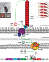
Model of curli biogenesis. Excluding CsgD, the master curli regulator, all Csg proteins have Sec-dependent signal sequences allowing their secretion into the periplasm. The lipoprotein CsgG forms a pore-like structure in the outer membrane. The major subunit protein CsgA and the nucleator CsgB are secreted to the cell surface in a CsgG- and CsgE-dependent manner. CsgF associates with the outer membrane and is required for cell association of the minor curli fiber subunit CsgB. Situated at the cell surface, CsgB nucleates soluble, unstructured CsgA into a highly ordered amyloid fiber. Curli production can be visualized by CR binding which is absent in a csgA mutant and by transmission electron microscopy (left inserts). Also shown are two CsgA subunits interacting in a cross-β conformation with the R1–R5 interaction depicted.
Figure 2
Figure 2
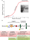
Schematic representation of amyloid polymerization. (a) Typical ThT fluorescence kinetics of soluble purified CsgA monomers polymerizing into curli [15]. The lag, growth and stationary phases are indicated. The blue arrow indicates the end of the lag phase. The insert shows a transmission electron micrograph of CsgA fibers formed when purified CsgA is allowed to polymerize in vitro as described by Wang et al. [8]. Scale bar: 500 nm. (b) The transition from soluble, monomeric proteins into polymeric and insoluble amyloid fibers is characterized by distinguishable steps that result in loss- or gain-of-function properties for the protein. In the lag phase, soluble protein assembles into a common intermediate or nucleus that is proposed to be toxic to membranes [29,88]. The formation of the intermediate is proposed to be the rate-limiting step of amyloidogenesis. Once the nucleus is formed, monomers are templated into growing amyloid fibers causing an increase in ThT fluorescence. When the monomer population is depleted, elongation stops and enters the stationary phase. The green boxes below the schematic highlight properties of some of the functional amyloids, whereas the red boxes highlight some general properties of disease-associated amyloids.

References

    1. Dobson CM. Structural biology: prying into prions. Nature. 2005;435:747–749. - PubMed
    1. Collinson SK, Parker JM, Hodges RS, Kay WW. Structural predictions of AgfA, the insoluble fimbrial subunit of Salmonella thin aggregative fimbriae. J Mol Biol. 1999;290:741–756. - PubMed
    1. Shewmaker F, McGlinchey RP, Thurber KR, McPhie P, Dyda F, Tycko R, Wickner RB. The functional curli amyloid is not based on in-register parallel {beta}-sheet structure. J Biol Chem. 2009;284:25065–25076. - PMC - PubMed
    1. Nordstedt C, Naslund J, Tjernberg LO, Karlstrom AR, Thyberg J, Terenius L. The Alzheimer A beta peptide develops protease resistance in association with its polymerization into fibrils. J Biol Chem. 1994;269:30773–30776. - PubMed
    1. Sitaras C, Naghavi M, Herrington MB. Sodium dodecyl sulfate-agarose gel electrophoresis for the detection and isolation of amyloid curli fibers. Anal Biochem. 2011;408:328–331. - PubMed

Publication types