Amyloid fibril formation by the glaucoma-associated olfactomedin domain of myocilin
- PMID: 22197377
- PMCID: PMC3323732
- DOI: 10.1016/j.jmb.2011.12.016
Amyloid fibril formation by the glaucoma-associated olfactomedin domain of myocilin
Abstract
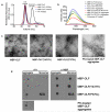
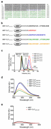
Myocilin is a protein found in the extracellular matrix of trabecular meshwork tissue, the anatomical region of the eye involved in regulating intraocular pressure. Wild-type (WT) myocilin has been associated with steroid-induced glaucoma, and variants of myocilin have been linked to early-onset inherited glaucoma. Elevated levels and aggregation of myocilin hasten increased intraocular pressure and glaucoma-characteristic vision loss due to irreversible damage to the optic nerve. In spite of reports on the intracellular accumulation of mutant and WT myocilin in vitro, cell culture, and model organisms, these aggregates have not been structurally characterized. In this work, we provide biophysical evidence for the hallmarks of amyloid fibrils in aggregated forms of WT and mutant myocilin localized to the C-terminal olfactomedin (OLF) domain. These fibrils are grown under a variety of conditions in a nucleation-dependent and self-propagating manner. Protofibrillar oligomers and mature amyloid fibrils are observed in vitro. Full-length mutant myocilin expressed in mammalian cells forms intracellular amyloid-containing aggregates as well. Taken together, this work provides new insights into and raises new questions about the molecular properties of the highly conserved OLF domain, and suggests a novel protein-based hypothesis for glaucoma pathogenesis for further testing in a clinical setting.
Copyright © 2011. Published by Elsevier Ltd.
Figures


Similar articles
-
Molecular Insights into Myocilin and Its Glaucoma-Causing Misfolded Olfactomedin Domain Variants.Acc Chem Res. 2021 May 4;54(9):2205-2215. doi: 10.1021/acs.accounts.1c00060. Epub 2021 Apr 13. Acc Chem Res. 2021. PMID: 33847483 Free PMC article. Review.
-
The glaucoma-associated olfactomedin domain of myocilin forms polymorphic fibrils that are constrained by partial unfolding and peptide sequence.J Mol Biol. 2014 Feb 20;426(4):921-35. doi: 10.1016/j.jmb.2013.12.002. Epub 2013 Dec 9. J Mol Biol. 2014. PMID: 24333014 Free PMC article.
-
The stability of myocilin olfactomedin domain variants provides new insight into glaucoma as a protein misfolding disorder.Biochemistry. 2011 Jul 5;50(26):5824-33. doi: 10.1021/bi200231x. Epub 2011 Jun 9. Biochemistry. 2011. PMID: 21612213 Free PMC article.
-
The glaucoma-associated olfactomedin domain of myocilin is a novel calcium binding protein.J Biol Chem. 2012 Dec 21;287(52):43370-7. doi: 10.1074/jbc.M112.408906. Epub 2012 Nov 5. J Biol Chem. 2012. PMID: 23129764 Free PMC article.
-
Myocilin misfolding and glaucoma: A 20-year update.Prog Retin Eye Res. 2023 Jul;95:101188. doi: 10.1016/j.preteyeres.2023.101188. Epub 2023 May 20. Prog Retin Eye Res. 2023. PMID: 37217093 Free PMC article. Review.
Cited by
-
Evidence for S331-G-S-L within the amyloid core of myocilin olfactomedin domain fibrils based on low-resolution 3D solid-state NMR spectra.bioRxiv [Preprint]. 2024 Aug 9:2024.08.09.606901. doi: 10.1101/2024.08.09.606901. bioRxiv. 2024. PMID: 39149386 Free PMC article. Preprint.
-
Molecular Insights into Myocilin and Its Glaucoma-Causing Misfolded Olfactomedin Domain Variants.Acc Chem Res. 2021 May 4;54(9):2205-2215. doi: 10.1021/acs.accounts.1c00060. Epub 2021 Apr 13. Acc Chem Res. 2021. PMID: 33847483 Free PMC article. Review.
-
A Novel Luciferase Assay For Sensitively Monitoring Myocilin Variants in Cell Culture.Invest Ophthalmol Vis Sci. 2016 Apr 1;57(4):1939-50. doi: 10.1167/iovs.15-18789. Invest Ophthalmol Vis Sci. 2016. PMID: 27092720 Free PMC article.
-
Competition between inside-out unfolding and pathogenic aggregation in an amyloid-forming β-propeller.Nat Commun. 2024 Jan 2;15(1):155. doi: 10.1038/s41467-023-44479-2. Nat Commun. 2024. PMID: 38168102 Free PMC article.
-
Structure‒function‒pathogenicity analysis of C-terminal myocilin missense variants based on experiments and 3D models.Front Genet. 2022 Oct 4;13:1019208. doi: 10.3389/fgene.2022.1019208. eCollection 2022. Front Genet. 2022. PMID: 36267417 Free PMC article.
References
-
- Alward WL. Medical management of glaucoma. N. Engl. J. Med. 1998;339:1298–307. - PubMed
-
- Kass MA, Heuer DK, Higginbotham EJ, Johnson CA, Keltner JL, Miller JP, Parrish RK, Wilson MR, Gordon MO. The Ocular Hypertension Treatment Study: a randomized trial determines that topical ocular hypotensive medication delays or prevents the onset of primary open-angle glaucoma. Archives of Ophthalmology. 2002;120:701–13. discussion 829-30. - PubMed
Publication types
MeSH terms
Substances
Grants and funding
LinkOut - more resources
Full Text Sources
Other Literature Sources
Medical

