. 2012 Mar;33(8):2482-91.
doi: 10.1016/j.biomaterials.2011.12.004. Epub 2011 Dec 23.

Mechanical derivation of functional myotubes from adipose-derived stem cells

Affiliations

Mechanical derivation of functional myotubes from adipose-derived stem cells

Yu Suk Choi et al. Biomaterials. 2012 Mar.

Abstract

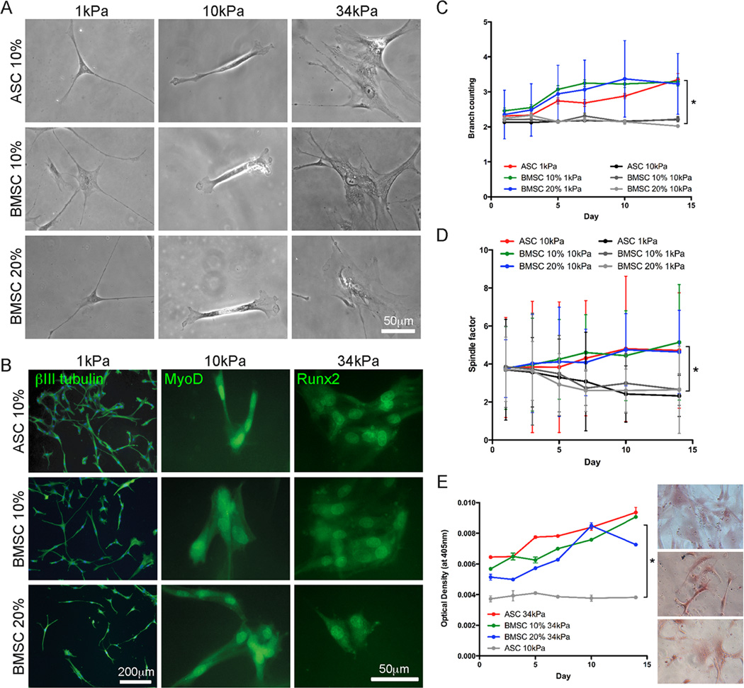
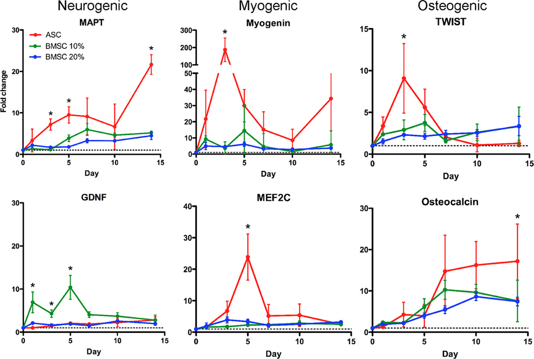
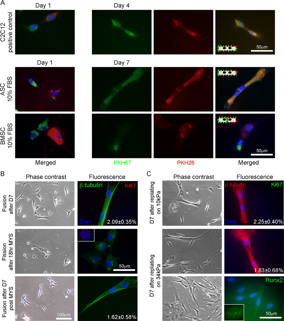
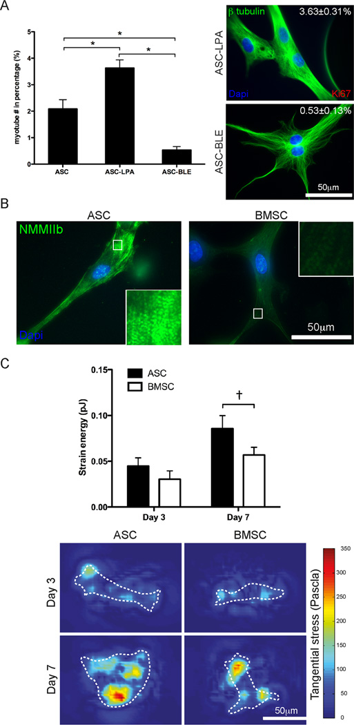
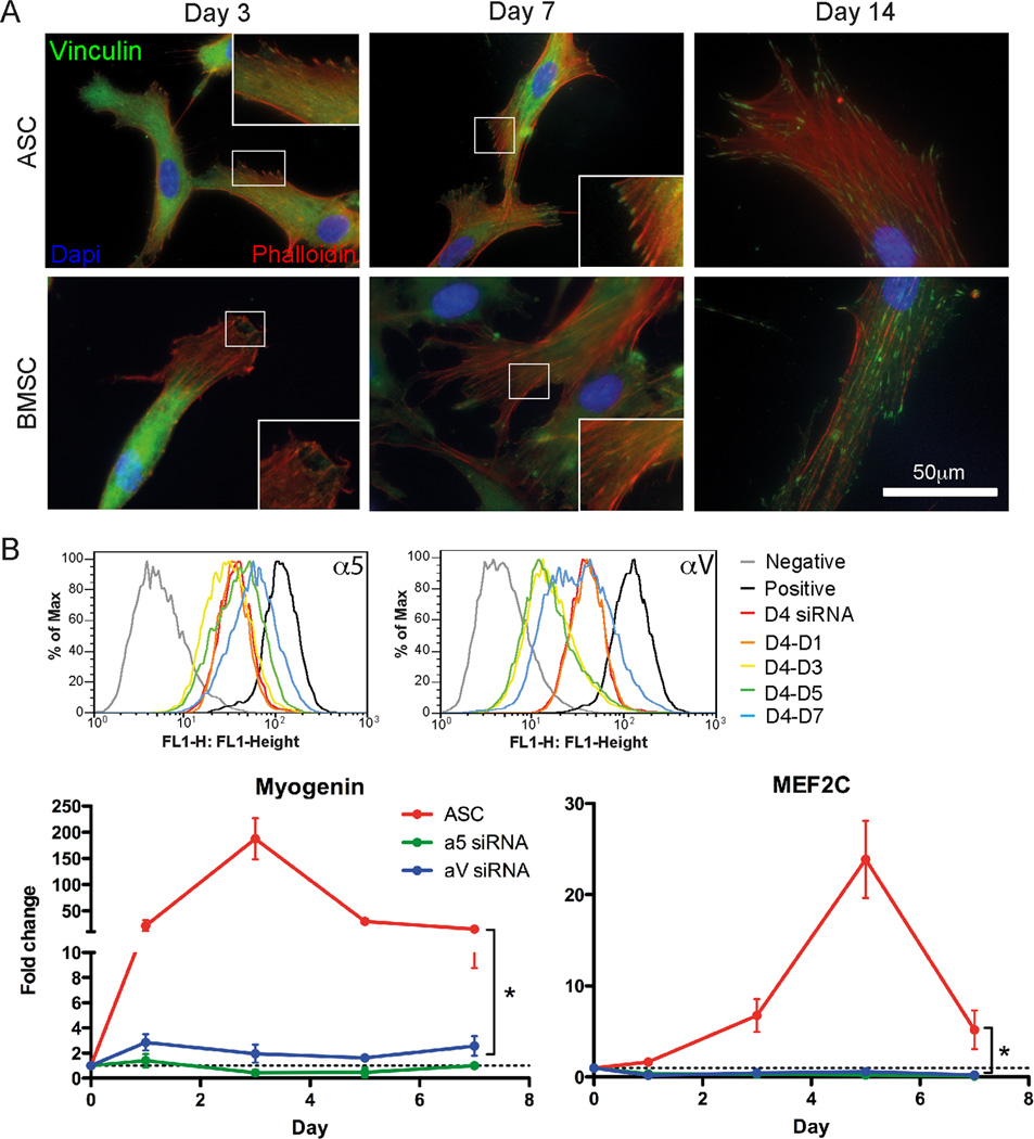
Though reduced serum or myoblast co-culture alone can differentiate adipose-derived stem cells (ASCs) into mesenchymal lineages, efficiency is usually not sufficient to restore function in vivo. Often when injected into fibrotic muscle, their differentiation may be misdirected by the now stiffened tissue. Here ASCs are shown to not just simply reflect the qualitative stiffness sensitivity of bone marrow-derived stem cells (BMSCs) but to exceed BMSC myogenic capacity, expressing the appropriate temporal sequence of muscle transcriptional regulators on muscle-mimicking extracellular matrix in a tension and focal adhesion-dependent manner. ASCs formed multi-nucleated myotubes with a continuous cytoskeleton that was not due to misdirected cell division; microtubule depolymerization severed myotubes, but after washout, ASCs refused at a rate similar to pre-treated values. BMSCs never underwent stiffness-mediated fusion. ASC-derived myotubes, when replated onto non-permissive stiff matrix, maintained their fused state. Together these data imply enhanced mechanosensitivity for ASCs, making them a better therapeutic cell source for fibrotic muscle.


Conflict of interest statement

Disclosure

The authors indicate no potential conflicts of interest.

Figures

Fig. 1
Fig. 1
ASC and BMSC differentiation on matrices with lineage-specific stiffness. (A) Representative morphology in phase contrast and (B) lineage-specific marker expression in fluorescence (βIII tubulin for neurogenic, MyoD for myogenic, and Runx2 for osteogenic lineages) in ASCs and BMSCs cultured in the indicated matrix and serum conditions after 1 week in culture. (C) The number of branches that ASCs and BMSCs formed over 2 weeks in culture in the indicated matrix and serum conditions was quantified. (D) The spindle-shaped morphology of muscle was determined by a spindle factor, the major axis/minor axis of cell, for ASCs and BMSCs over 2 weeks in culture in the indicated matrix and serum conditions. (E) ASCs and BMSCs were stained for Alizarin Red S mineralization over 2 weeks in culture in the indicated matrix and serum conditions. Inset images are representative of ASCs (top) and BMSCs (10 and 20% serum, middle and bottom, respectively) cultured on the 34 kPa matrix. All data shown in mean ± standard deviation for triplicate experiments. *P < 0.05.
Fig. 2
Fig. 2
Lineage-specific mRNA is highest in ASCs. Gene expression of MAPT, GDNF, Myogenin, MEF2C, TWIST, and Osteocalcin was assessed by qPCR. Lineages are indicated at the top of each column. Expression was monitored as a function of time for ASCs cultured in 10% serum-containing media (red) and BMSCs cultured in either 10 (green) or 20% serum-containing media (blue). The dashed line indicates expression in the undifferentiated cell to which all data is normalized. * indicated P < 0.05.
Fig. 3
Fig. 3
Multi-nucleated cell formation in ASCs on myogenic matrices. (A) PKH67 (green) and PKH26 (red) labeled C2C12 myoblasts (top), ASCs (middle), and BMSCs (bottom) were mixed with their oppositely labeled cell of the same type, plated on 10kPa matrix, and monitored for dye transfer between cells. After 1 day, all three cell sources did not exhibit transfer between adjacent cells (left). Transfer between adjacent myotubes occurred by day4, between adjacent ASCs by day 7, and never occurred for BMSCs (right). The inset schematics illustrate this process. (B) ASCs were also examined by β tubulin (green, continuous cytoskeleton) and Ki67 (red, proliferation) after day 7. The percent of bi-nucleated cells is inset in the fluorescent image (top). Myoseverin was added to cells after day 7, reverting bi-nucleated cells to singly-nucleated cells (middle), with some Ki67 positive staining (inset). After washout, ASCs refused at a rate similar to the pre-treated cells as indicated (bottom). (C) After 7 days in culture on 10kPa matrix, ASCs were replated onto either 10 (top) or 34 kPa matrices (middle and bottom) for an additional 7 days. The percent of bi-nucleated cells is inset in the fluorescent images stained for β tubulin (red) and ki67(green). Singly-nucleated cells replated from 10 to 34 kPa were also stained for Runx2 (green, bottom and inset).
Fig. 4
Fig. 4
Mechanotransduction in ASCs and BMSCs regulates differentiation. (A) Myotube frequency was increased by the contractile agonist Lysophosphatidic acid (LPA) and decreased by the NMMII inhibitor Blebbistatin (BLE). *P < 0.001. (B) Immunofluorescent staining of NMMIIb in ASCs and BMSCs with higher magnification images inset to illustrate striation assembly. (C) Strain energy was measured by TFM (top). Representative images of the tangential stress (bottom) generated from TFM are shown at both days 3 and 7 with white cell outlines. The colormap indicates the magnitude of the stress as measured in Pascals. Data is reported as the mean ± standard error from triplicate experiments representing greater than 12 cells. P = 0.07.
Fig. 5
Fig. 5
Mechanical induction is differentially transduced by intracellular signals via focal adhesions. (A) Assembled vinculin adhesions (green) were found in ASCs (left) at day 3 and in BMSCs (right) by day 7. F-actin (red phalloidin) and nuclei (blue) are also shown. (B) Flow cytometry plots of ASCs transfected with siRNA for α5 (top)or αV integrin (bottom) on TCP for 4 days (D4 siRNA) or ASCs that were also then replated on myogenic 10kPa matrices for the indicate time in days (left). qPCR analysis of myogenic markers Myogenin and MEF2C showed total inhibition for siRNA-treated cells of either integrin. Data represents the mean ± standard deviation of triplicate experiments. *P < 0.05.

References

    1. Stedman HH, Sweeney HL, Shrager JB, Maguire HC, Panettieri RA, Petrof B, et al. The mdx mouse diaphragm reproduces the degenerative changes of Duchenne muscular dystrophy. Nature. 1991;352:536–539. - PubMed
    1. Bushby K, Finkel R, Birnkrant DJ, Case LE, Clemens PR, Cripe L, et al. Diagnosis and management of Duchenne muscular dystrophy, part 1: diagnosis, and pharmacological and psychosocial management. Lancet Neurol. 2010;9:77–93. - PubMed
    1. Davies JE, Rose C, Sarkar S, Rubinsztein DC. Cystamine suppresses polyalanine toxicity in a mouse model of oculopharyngeal muscular dystrophy. Sci Transl Med. 2010;2 34ra40. - PubMed
    1. Engler AJ, Sen S, Sweeney HL, Discher DE. Matrix elasticity directs stem cell lineage specification. Cell. 2006;126:677–689. - PubMed
    1. Breitbach M, Bostani T, Roell W, Xia Y, Dewald O, Nygren JM, et al. Potential risks of bone marrow cell transplantation into infarcted hearts. Blood. 2007;110:1362–1369. - PubMed

Publication types

MeSH terms