. 2012 Apr 1;229(1):48-56.
doi: 10.1016/j.bbr.2011.12.019. Epub 2011 Dec 20.

Expression of HIV-Tat protein is associated with learning and memory deficits in the mouse

Affiliations

Expression of HIV-Tat protein is associated with learning and memory deficits in the mouse

Amanda N Carey et al. Behav Brain Res. .

Abstract

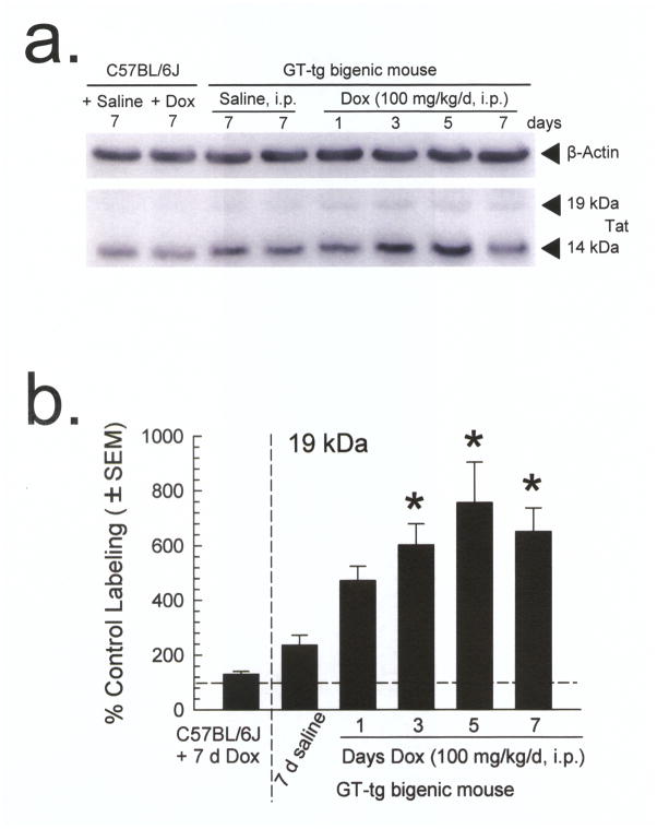
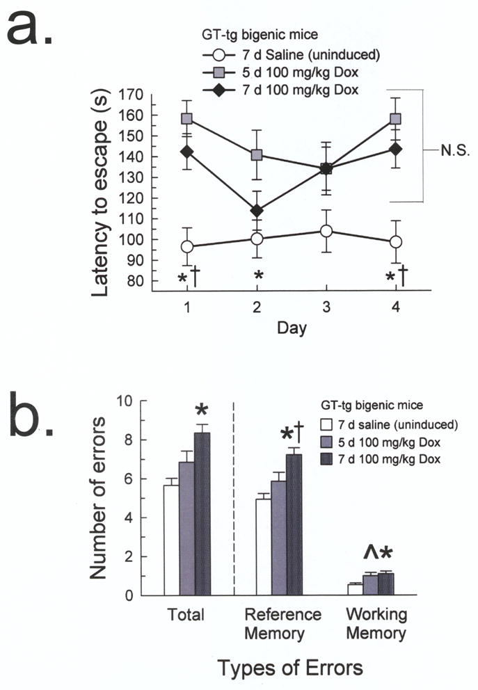
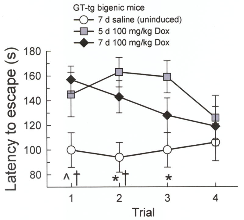
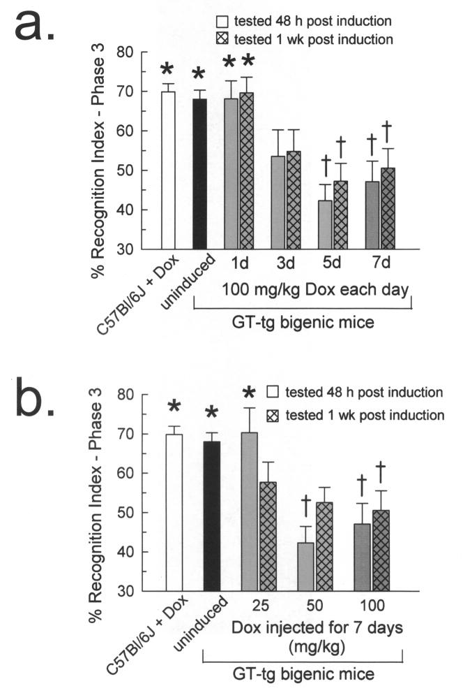
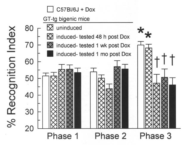
HIV-Tat protein has been implicated in the pathogenesis of HIV-1 neurological complications (i.e., neuroAIDS), but direct demonstrations of the effects of Tat on behavior are limited. GT-tg mice with a doxycycline (Dox)-inducible and brain-selective tat gene coding for Tat protein were used to test the hypothesis that the activity of Tat in brain is sufficient to impair learning and memory processes. Western blot analysis of GT-tg mouse brains demonstrated an increase in Tat antibody labeling that seemed to be dependent on the dose and duration of Dox pretreatment. Dox-treated GT-tg mice tested in the Barnes maze demonstrated longer latencies to find an escape hole and displayed deficits in probe trial performance versus uninduced GT-tg littermates, suggesting Tat-induced impairments of spatial learning and memory. Reversal learning was also impaired in Tat-induced mice. Tat-induced mice additionally demonstrated long-lasting (up to one month) deficiencies in novel object recognition learning and memory performance. Furthermore, novel object recognition impairment was dependent on the dose and duration of Dox exposure, suggesting that Tat exposure progressively mediated deficits. These experiments provide evidence that Tat protein expression is sufficient to mediate cognitive abnormalities seen in HIV-infected individuals. Moreover, the genetically engineered GT-tg mouse may be useful for improving our understanding of the neurological underpinnings of neuroAIDS-related behaviors.


Figures

Fig. 1
Fig. 1. Doxycycline-induced HIV-1 Tat protein expression in GT-tg mouse brain
(a) Representative Western blots from whole brain. The β-actin antibody labeled a single band (upper panel) corresponding to the weight of the β-actin protein of similar intensity across all samples. By contrast, the Tat antibody labeled a number of proteins non-selectively (lower panel), but only demonstrated a difference in labeling intensity that corresponded to the presence and duration of Dox administered. (b) Summary graph of the quantified whole brain Western blots. All band intensities are plotted as percent control (saline-treated C57BL/6J) labeling. Dox treatment resulted in an increase in Tat-antibody labeling in GT-tg mice dependent on the duration of pretreatment. Labeling intensity differences corresponded to the presence and duration of Dox administered at 19 kDa, which has been suggested to be the observed weight of expressed Tat protein in the GT-tg mouse.
Fig. 2
Fig. 2. Tat-induced mice demonstrated longer escape latencies and committed more Barnes maze acquisition errors versus uninduced GT-tg mice
(a) The 5 day Tat-induced mice (100 mg/kg Dox, i.p.; gray squares) demonstrated longer escape latencies compared to uninduced GT-tg mice (0.9% saline, i.p., 7 days; white circles) on days 1, 2, and 4, while the 7 day Tat-induced mice (100 mg/kg Dox, i.p.; black diamonds) demonstrated longer latencies on days 1 and 4. (n = 9–18; n.s. = not significant, * = different from 5 day Tat-induced mice, † = different from 7 day Tat-induced mice, p < 0.05, Tukey’s HSD). (b) The 7 day Tat-induced mice (dark gray patterned bars) committed more total, reference memory, and working memory errors than the uninduced mice (white bars). The 5 day Tat-induced mice (gray bars) showed a trend toward committing more working memory errors than the uninduced mice. (n= 8–16; ^ = trend from uninduced mice, p = 0.06, * = different from uninduced mice, † = different from 5 day Tat-induced mice, p < 0.05, Tukey’s HSD)
Fig. 3
Fig. 3. Tat-induced mice required more trials to learn a new escape location during a Barnes maze reversal learning task
Escape latencies did not differ between the GT-tg mice induced to express Tat with Dox (100 mg/kg, i.p.) for 5 days (gray squares) or 7 days (black diamonds). The 5 day Tat-induced mice demonstrated longer escape latencies compared to uninduced GT-tg mice (0.9% saline, i.p., 7 days) (white circles) on trials 2 and 3, and showed a trend on trial 1. The 7 day Tat-induced mice demonstrated longer escape latencies compared to uninduced mice on trials 1 and 2. (n = 9–19; ^ = trend from 5 day Tat-induced mice, p = 0.11, * = different from 5 day Tat-induced mice, † = different from 7 day Tat-induced mice, p < 0.05, Tukey’s HSD).
Fig. 4
Fig. 4. Tat-induced GT-tg mice exhibited phase 3 novel object recognition deficits
GT-tg mice were administered the maximum Dox dose (100 mg/kg, i.p.) for 1, 3, 5, or 7 days (a, gray bars) or 25, 50, or 100 mg/kg Dox for 7 days (b, gray bars). Groups were tested 2 days (solid bars) or 7 days (hatched bars) after induction. (a) Tat-induced GT-tg mice demonstrated a Dox duration-dependent deficit in phase 3 novel object recognition. The 5 and 7 day Tat-induced mice showed differences in phase 3 RI when compared to C57BL/6J mice and uninduced littermates. (n = 17–53 mice/bar; * = different from matching phase 1 response, † = different from phase 3 response of C57BL/6J and uninduced mice, p ≤ 0.05, Tukey’s HSD). (b) Tat-induced GT-tg mice demonstrated a duration-dependent deficit in phase 3 novel object recognition. Mice induced for 7 days with 50 or 100 mg/kg Dox were impaired in phase 3 NOR. Mice induced with 25 mg/kg Dox and tested one week after induction also demonstrated impairment in phase 3. (n = 17–53 mice/bar; * = different from matching phase 1 response, † = different from phase 3 response of C57BL/6J and uninduced mice, p ≤ 0.05, Tukey’s HSD).
Fig. 5
Fig. 5. Tat-induced GT-tg mice exhibited persistent suppression of phase 3 novel object recognition performance
Mice were induced to express Tat with 100 mg/kg Dox for 7 days and tested 2 days (light gray thatched bars), 7 days (dark gray thatched bars), or 30 days (black bars) after the completion of induction. Phase 3 NOR of the C57BL/6J and uninduced mice was not different. Tat-induced mice tested at all time points spent less time on the novel object in phase 3 than controls and showed no increase in phase 3 RIs from phase 1. (n = 17–53 mice/bar; * = significant increase from phase 1 response, † = different from phase 3 response of C57BL/6J and uninduced mice, p < 0.05, Tukey’s HSD).

References

    1. Frankel AD, Young JA. HIV-1 fifteen proteins and an RNA. Ann Rev Biochem. 1998;67:1–25. - PubMed
    1. Power C, McArthur JC, Nath A, Wehrly K, Mayne M, Nishio J, Langelier T, Johnson RT, Chesebro B. Neuronal death induced by brain-derived human immunodeficiency virus type 1 envelope genes differs between demented and nondemented AIDS patients. J Virol. 1998;72:9045–9053. - PMC - PubMed
    1. Brack-Werner R. Astrocytes: HIV cellular reservoirs and important participants in neuropathogenesis. AIDS. 1999;13:1–22. - PubMed
    1. Johnston JB, Zhang K, Silva C, Shalinsky DR, Conant K, Ni W, Corbett D, Yong VW, Power C. HIV-1 Tat neurotoxicity is prevented by matrix metalloproteinase inhibitors. Ann Neurol. 2001;49:230–241. - PubMed
    1. Glowa JR, Panlilio LV, Brenneman DE, Gozes I, Fridkin M, Hill JM. Learning impairment following intracerebral administration of the HIV envelope protein gp120 or a VIP antagonist. Brain Res. 1992;570:49–53. - PubMed

Publication types

MeSH terms