. 2011 Dec 25;15(2):197-204.
doi: 10.1038/nn.3019.

Encapsulated therapeutic stem cells implanted in the tumor resection cavity induce cell death in gliomas

Affiliations

Encapsulated therapeutic stem cells implanted in the tumor resection cavity induce cell death in gliomas

Timo M Kauer et al. Nat Neurosci. .

Abstract

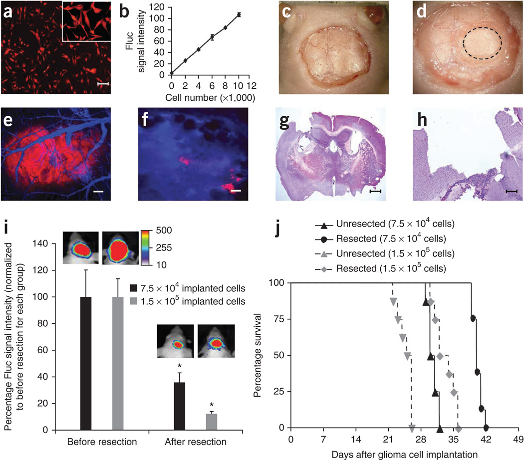
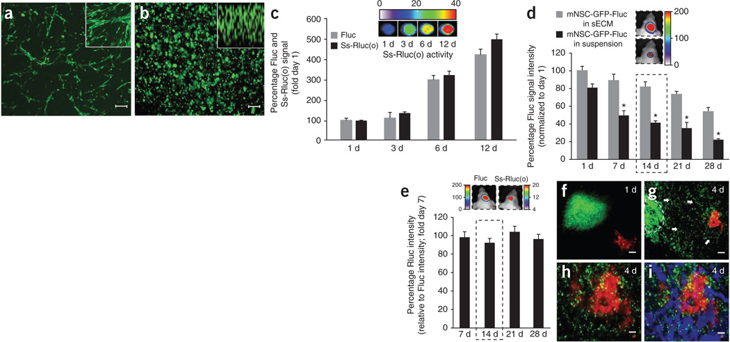
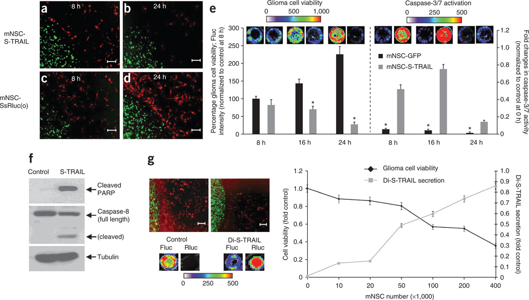
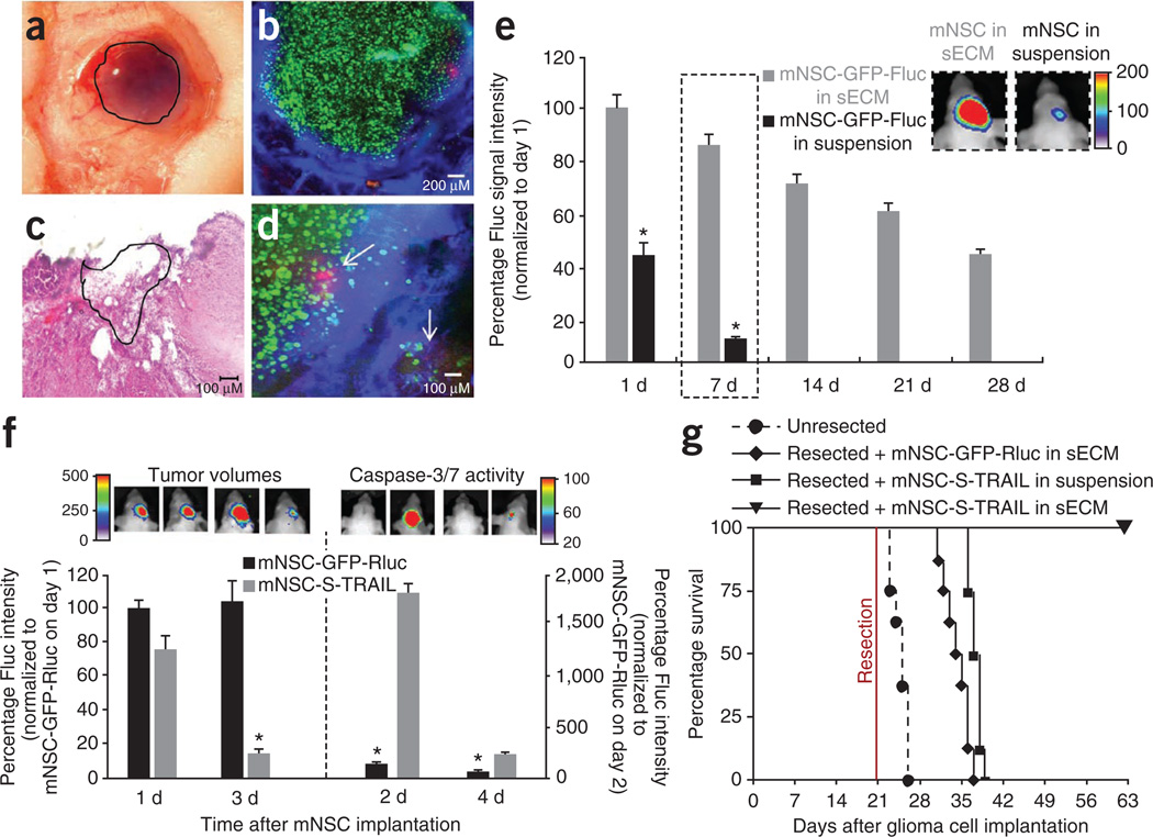
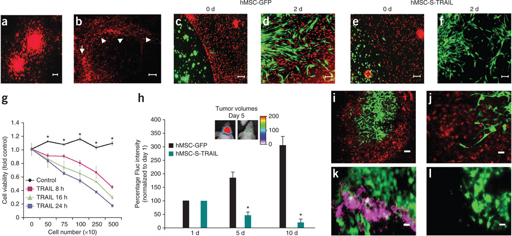
Therapeutically engineered stem cells have shown promise for glioblastoma multiforme (GBM) therapy; however, key preclinical studies are urgently needed for their clinical translation. In this study, we investigated a new approach to GBM treatment using therapeutic stem cells encapsulated in biodegradable, synthetic extracellular matrix (sECM) in mouse models of human GBM resection. Using multimodal imaging, we first showed quantitative surgical debulking of human GBM tumors in mice, which resulted in increased survival. Next, sECM encapsulation of engineered stem cells increased their retention in the tumor resection cavity, permitted tumor-selective migration and release of diagnostic and therapeutic proteins in vivo. Simulating the clinical scenario of GBM treatment, the release of tumor-selective S-TRAIL (secretable tumor necrosis factor apoptosis inducing ligand) from sECM-encapsulated stem cells in the resection cavity eradicated residual tumor cells by inducing caspase-mediated apoptosis, delayed tumor regrowth and significantly increased survival of mice. This study demonstrates the efficacy of encapsulated therapeutic stem cells in mouse models of GBM resection and may have implications for developing effective therapies for GBM.


Figures

Figure 1
Figure 1. Tumor resection prolongs survival of mice bearing GBM
(a,b) Cell transduction. Human U87 GBM cells were transduced with LV-Fluc-mCherry, and cells were imaged 48 h later for mCherry expression and Fluc activity. Photomicrograph of U87 cells expressing Fluc-mCherry (a) and plot revealing the correlation between U87-Fluc-mCherry cell number and Fluc activity (b). (cf) Cranial implantation. A cranial window was established in mice and U87-Fluc-mCherry cells (7.5 × 10⁴ or 1.5 × 10⁵) were implanted in the cranial window. Light images of the mouse skull with skin removed (c), drilled rim around the cranial window (d). Dashed circle indicates the tumor growing area in the cranial window. (e,f) IVM. Mice with established U87-Fluc-mCherry GBMs in the cranial window were injected with a blood-pool agent, AngioSense-750. Photomicrographs before (e) and after (f) tumor resection (red, tumor; blue, vasculature). (g,h) Photomicrographs of low (g) and high (h) magnification hematoxylin and eosin staining of brain sections showing tumor resection cavity. (i) Plot of the Fluc signal intensity and representative visible light plus superimposed bioluminescence images (color scale units, photons min−1 cm−2; here and in subsequent figures) before and after tumor resection in mice implanted with 7.5 × 10⁴ (resected 14 d after implantation) or 1.5 × 10⁵ (resected 21 d after implantation) GBM cells. *P < 0.05 versus before resection for each group. Data are mean ± s.e.m. (j) Kaplan-Meier survival curves of mice with and without resected U87-Fluc-mCherry tumors. P < 0.05 resected versus un-resected tumors for each group. Scale bars, 100 µm (a,e,f,h) and 400 µm (g). Original magnifications: ×2 (c) and ×4 (d).
Figure 2
Figure 2. Characterizing engineered mNSCs in biocompatible sECM in vitro and in vivo
(a,b) Photomicrographs of mNSCs expressing GFP-Fluc grown in monolayers, with higher magnification in inset (a) and encapsulated in sECM, with z-stack in inset (b). (c) Plot of cell proliferation and protein secretion and representative Rluc images for mNSCs coexpressing GFP-Fluc and Ss-Rluc(o), encapsulated in sECM and followed by simultaneous Fluc and Rluc imaging of cells and culture medium, respectively. (d) mNSC-GFP-Fluc cell survival in the brain over a period of 4 weeks when implanted in sECM versus in suspension. Mice were imaged serially for mNSC survival by Fluc activity. Representative images from day 14 mice (dashed outline) are shown. *P < 0.05 versus unencapsulated mNSCs. (e) Ratio of Rluc signal intensity relative to Fluc signal intensity. mNSCs expressing GFP-Fluc plus Ss-Rluc(o) were encapsulated in sECM and implanted intracranially, and cell viability (Fluc signal) and protein secretion (Rluc signal) were followed by simultaneous Fluc and Rluc imaging in vivo. Representative images from day 14 mice (dashed outline) are shown. (fi) IVM images showing mNSCs (green) and tumor cells (red) on day 1 (f) and day 4 (gi) after mNSC implantation. Mice bearing U87-mCherry-Fluc GBMs in the cranial windows were implanted with mNSC-GFP-Rluc cells encapsulated in sECM, 1 mm away from the established tumor. Dashed line, encapsulated mNSCs; arrows, mNSC migration in g; blue in i, tumor vasculature. Scale bars: 100 µm (a,b,h,i) and 200 µm (f,g). Original magnifications: ×20 (a,b insets). Data are mean ± s.e.m.
Figure 3
Figure 3. mNSCs expressing therapeutic S-TRAIL induce GBM cell death in vitro
(ae) mNSCs (green) expressing Ss-Rluc(o) or S-TRAIL were encapsulated in sECM and placed in a culture dish containing human GBM U87-Fluc-mCherry cells (red). Photomicrographs show sECM-encapsulated mNSCs at 8 h (a,c) and 24 h (b,d). Plot shows tumor cell viability (*P < 0.05 versus controls) and caspase-3/7 activation (*P < 0.05 versus mNSC-S-TRAIL) over 24 h when cultured with either sECM-encapsulated mNSC-GFP or mNSC-S-TRAIL cells (e). (f) Western blot analysis on GBM cells collected 8 h after sECM-encapsulated mNSC-S-TRAIL or mNSC-GFP (control) cell placement in the culture dish (see Supplementary Fig. 4 for uncropped blots). (g) Representative images and summary graph demonstrating the effect of the release of Di-S-TRAIL from sECM-encapsulated mNSCs cultured with U87-mCherry-Fluc cells at increasing ratios of stem cell to tumor cell. After 24 h of culture, fluorescence photomicrographs (top left) were taken, Di-S-TRAIL was visualized by Rluc bioluminescence imaging and tumor cell viability was visualized by Fluc bioluminescence imaging. Scale bars, 100 µm. Data are mean ± s.e.m.
Figure 4
Figure 4. sECM-encapsulated mNSC-S-TRAIL cells transplanted into the tumor resection cavity increase survival of mice
(a) Light photomicrograph of the resection cavity containing sECM-encapsulated mNSCs (outlined area). (bd) mNSC-GFP-Fluc cells encapsulated in sECM were implanted intracranially in the resection cavity, and the mice were injected with Angiosense-750 (blue) intravenously and imaged by IVM and by serial Fluc bioluminescence imaging. (b) Fluorescence photomicrograph showing mNSCs (green) targeting residual GBM cells (red) in a tumor resection cavity with leaky vasculature (blue). (c) Hematoxylin and eosin image of sECM-encapsulated mNSC-GFP-Fluc cells implanted (outlined area) in the resection cavity. (d) Higher magnification fluorescence photomicrograph showing mNSCs (green) targeting residual GBM cells (red) indicated by arrows in a tumor resection cavity with leaky vasculature (blue). (e) Plot and representative images from day 7 (dashed outlines) of the relative mean Fluc signal intensity of mNSC-GFP-Fluc cells in suspension or encapsulated in sECM, placed in the GBM resection cavity (*P < 0.05 versus encapsulated mNSCs). (f,g) mNSC-S-TRAIL or mNSC-GFP-Rluc cells encapsulated in sECM or mNSC-S-TRAIL cells in suspension were implanted intracranially in the tumor resection cavity. (f) TRAIL-mediated caspase-3/7 activation and tumor volumes as assessed by serial bioluminescence imaging after aminoluciferin and luciferin injections, respectively. Caspase-3/7 activity, *P < 0.05 versus mNSC-S-TRAIL; tumor volumes, *P < 0.05 versus controls. (g) Kaplan-Meier survival curves. P < 0.05, resected + GFP-Rluc cells in sECM versus un-resected; P < 0.05, resected + mNSC-S-TRAIL cells in sECM versus resected + mNSC-S-TRAIL cells in suspension. Scale bars, 200 µm (b) and 100 µm (c,d). Original magnification, ×4 (a). Data are mean ± s.e.m.
Figure 5
Figure 5. sECM-encapsulated therapeutic human MSCs have anti-tumor effects on primary invasive human GBMs in vitro and in vivo
(a,b) Primary invasive GBM8-mCherry-Fluc cells grown as neurospheres in a collagen matrix (a) and brain section of mice bearing GBM8-mCherry-Fluc tumors, showing the highly invasive nature of GBM8 (b). Arrow, site of implantation; arrowheads, path of invasion. (cg) hMSCs expressing GFP or S-TRAIL were encapsulated in sECM and placed in a culture dish containing human GBM8-Fluc-mCherry cells. hMSCs (green) were followed for migration out of sECM, and GBM8 cells (red) were followed for their response to S-TRAIL secreted by hMSCs. Photomicrographs show sECM-encapsulated hMSCs on the day of plating (c,e) and 48 h after plating (d,f). (g) GBM8 cell viability at different time points after culturing with varying numbers of either sECM-encapsulated hMSC-GFP (control) or hMSC-S-TRAIL (TRAIL) cells. *P < 0.05 versus TRAIL at 8 h, 16 h and 24 h. (hj) Encapsulated hMSC-S-TRAIL or hMSC-GFP cells in sECM were implanted intracranially in the tumor resection cavity of mice bearing GBM8-mCherry-Fluc cells and mice were followed for changes in tumor volume by serial Fluc bioluminescence imaging and correlative immunohistochemistry. Plot and representative images show the relative mean Fluc signal intensity from mice bearing sECM-encapsulated hMSC-GFP or hMSC-S-TRAIL cells. *P < 0.05 versus control (h). (i,j) Low-magnification (i) and high-magnification (j) photomicrographs of serial brain sections of mice showing hMSCs (green) on day 5 after hMSC implantation in the GBM8 (red) resection cavity. (k,l) Representative images showing cleaved caspase-3 staining (purple) on brain sections from mice implanted with hMSC-S-TRAIL cells (green, k) and control cells (green, l) 5 d after treatment. Scale bars: 100 µm (a,cf,i), 200 µm (b) and 50 µm (jl). Data are mean ± s.e.m.

Comment in

Similar articles

Cited by

References

    1. Adamson C, et al. Glioblastoma multiforme: a review of where we have been and where we are going. Expert Opin. Investig. Drugs. 2009;18:1061–1083. - PubMed
    1. Affronti ML, et al. Overall survival of newly diagnosed glioblastoma patients receiving carmustine wafers followed by radiation and concurrent temozolomide plus rotational multiagent chemotherapy. Cancer. 2009;115:3501–3511. - PubMed
    1. Wen PY, Kesari S. Malignant gliomas in adults. N. Engl. J. Med. 2008;359:492–507. - PubMed
    1. Asthagiri AR, Pouratian N, Sherman J, Ahmed G, Shaffrey ME. Advances in brain tumor surgery. Neurol. Clin. 2007;25:975–1003. viii–ix. - PubMed
    1. Erpolat OP, et al. Outcome of newly diagnosed glioblastoma patients treated by radiotherapy plus concomitant and adjuvant temozolomide: a long-term analysis. Tumori. 2009;95:191–197. - PubMed

Publication types

MeSH terms