

Cross-talk between calcium signalling and protein phosphorylation at the thylakoid
- PMID: 22197893
- PMCID: PMC3970089
- DOI: 10.1093/jxb/err403
Cross-talk between calcium signalling and protein phosphorylation at the thylakoid
Abstract
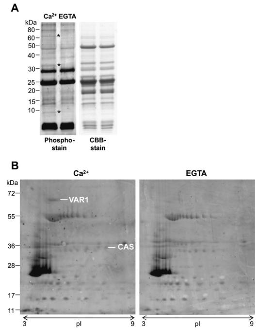
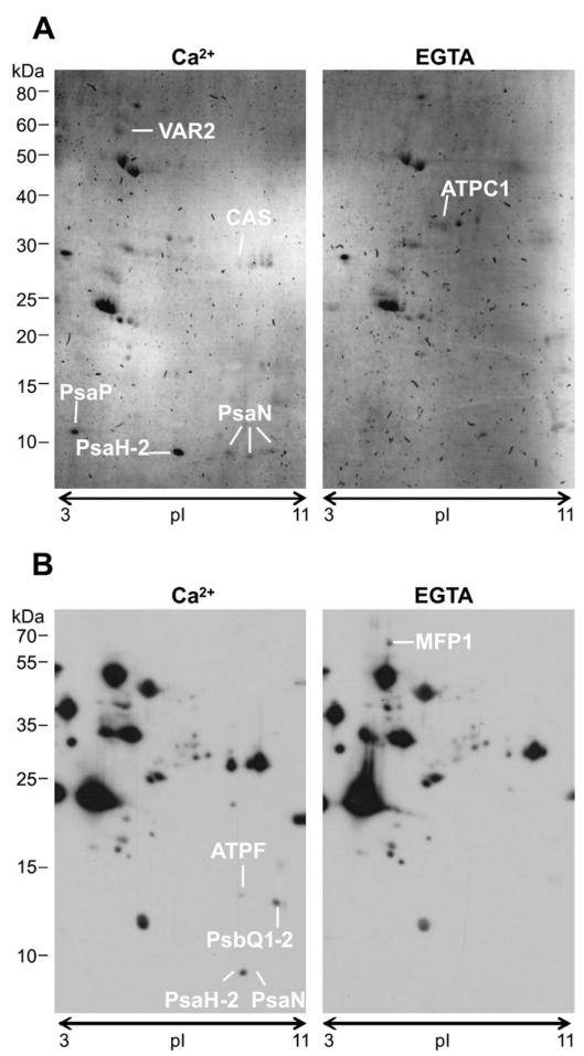
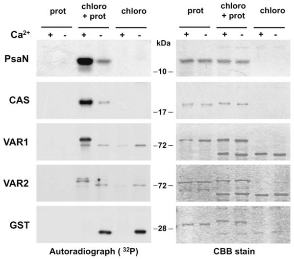
The role of protein phosphorylation for adjusting chloroplast functions to changing environmental needs is well established, whereas calcium signalling in the chloroplast is only recently becoming appreciated. The work presented here explores the potential cross-talk between calcium signalling and protein phosphorylation in chloroplasts and provides the first evidence for targets of calcium-dependent protein phosphorylation at the thylakoid membrane. Thylakoid proteins were screened for calcium-dependent phosphorylation by 2D gel electrophoresis combined with phospho-specific labelling and PsaN, CAS, and VAR1, among other proteins, were identified repeatedly by mass spectrometry. Subsequently their calcium-dependent phosphorylation was confirmed in kinase assays using the purified proteins and chloroplast extracts. This is the first report on the protein targets of calcium-dependent phosphorylation of thylakoid proteins and provides ground for further studies in this direction.
Figures
Similar articles
-
Correlation between spatial (3D) structure of pea and bean thylakoid membranes and arrangement of chlorophyll-protein complexes.BMC Plant Biol. 2012 May 25;12:72. doi: 10.1186/1471-2229-12-72. BMC Plant Biol. 2012. PMID: 22631450 Free PMC article.
-
Chloroplast biogenesis. Regulation of lipid transport to the thylakoid in chloroplasts isolated from expanding and fully expanded leaves of pea.Plant Physiol. 2001 Sep;127(1):184-93. doi: 10.1104/pp.127.1.184. Plant Physiol. 2001. PMID: 11553746 Free PMC article.
-
Chloroplast Ca2+ Fluxes into and across Thylakoids Revealed by Thylakoid-Targeted Aequorin Probes.Plant Physiol. 2018 May;177(1):38-51. doi: 10.1104/pp.18.00027. Epub 2018 Mar 20. Plant Physiol. 2018. PMID: 29559589 Free PMC article.
-
Evidence for nucleotide-dependent processes in the thylakoid lumen of plant chloroplasts--an update.FEBS Lett. 2012 Aug 31;586(18):2946-54. doi: 10.1016/j.febslet.2012.07.005. Epub 2012 Jul 13. FEBS Lett. 2012. PMID: 22796491 Review.
-
Light, redox state, thylakoid-protein phosphorylation and signaling gene expression.Trends Biochem Sci. 2003 Sep;28(9):467-70. doi: 10.1016/S0968-0004(03)00173-7. Trends Biochem Sci. 2003. PMID: 13678955 Review.
Cited by
-
A comparative study on photosynthetic characteristics and flavonoid metabolism between Camellia petelotii (Merr.) Sealy and Camellia impressinervis Chang &Liang.Front Plant Sci. 2022 Nov 24;13:1071458. doi: 10.3389/fpls.2022.1071458. eCollection 2022. Front Plant Sci. 2022. PMID: 36544877 Free PMC article.
-
Chloroplast Calcium Signaling in the Spotlight.Front Plant Sci. 2020 Mar 11;11:186. doi: 10.3389/fpls.2020.00186. eCollection 2020. Front Plant Sci. 2020. PMID: 32226434 Free PMC article. Review.
-
The role of calcium in chloroplasts--an intriguing and unresolved puzzle.Protoplasma. 2012 Oct;249(4):957-66. doi: 10.1007/s00709-011-0373-3. Epub 2012 Jan 8. Protoplasma. 2012. PMID: 22227834 Review.
-
Understanding the roles of the thylakoid lumen in photosynthesis regulation.Front Plant Sci. 2013 Oct 31;4:434. doi: 10.3389/fpls.2013.00434. Front Plant Sci. 2013. PMID: 24198822 Free PMC article. Review.
-
CsCIPK11-Regulated Metalloprotease CsFtsH5 Mediates the Cold Response of Tea Plants.Int J Mol Sci. 2023 Mar 27;24(7):6288. doi: 10.3390/ijms24076288. Int J Mol Sci. 2023. PMID: 37047263 Free PMC article.
References
-
- Aro EM, Suorsa M, Rokka A, Allahverdiyeva Y, Paakkarinen V, Saleem A, Battchikova N, Rintamaki E. Dynamics of photosystem II: a proteomic approach to thylakoid protein complexes. Journal of Experimental Botany. 2005;56:347–356. - PubMed
-
- Baginsky S, Tiller K, Pfannschmidt T, Link G. PTK, the chloroplast RNA polymerase-associated protein kinase from mustard (Sinapis alba), mediates redox control of plastid in vitro transcription. Plant Molecular Biology. 1999;39:1013–1023. - PubMed
Publication types
MeSH terms
Substances
Grants and funding
LinkOut - more resources
Full Text Sources
Molecular Biology Databases
