
B cell-helper neutrophils stimulate the diversification and production of immunoglobulin in the marginal zone of the spleen

Irene Puga et al. Nat Immunol. .

Erratum in

  • Nat Immunol. 2014 Feb;15(2):205

Abstract

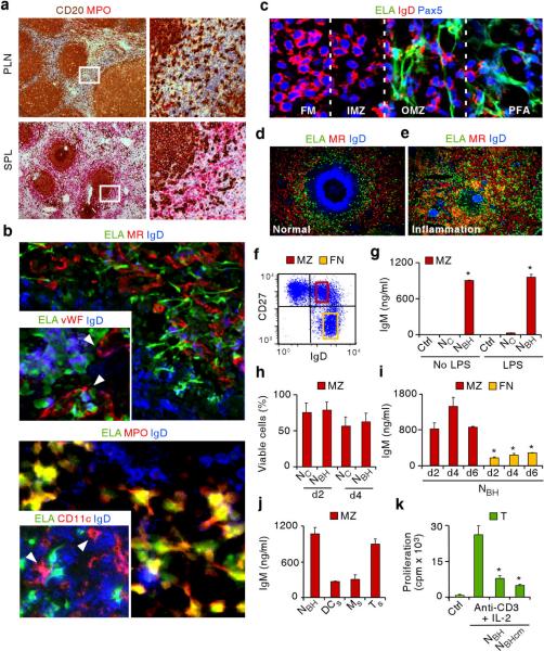
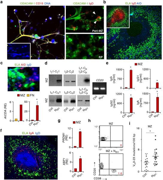
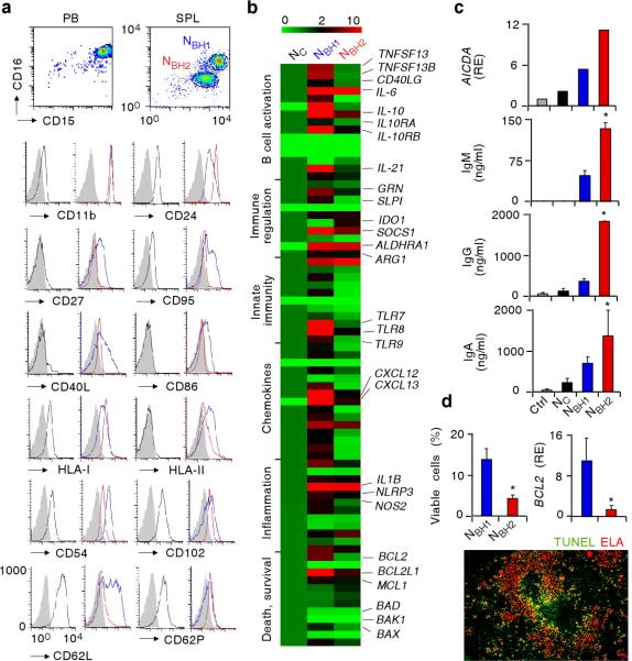
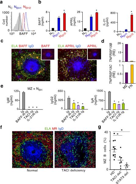
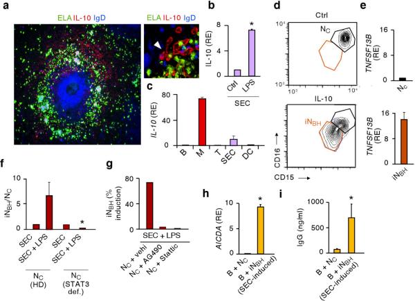
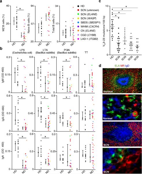
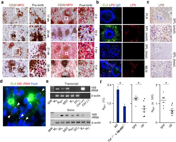
Neutrophils use immunoglobulins to clear antigen, but their role in immunoglobulin production is unknown. Here we identified neutrophils around the marginal zone (MZ) of the spleen, a B cell area specialized in T cell-independent immunoglobulin responses to circulating antigen. Neutrophils colonized peri-MZ areas after postnatal mucosal colonization by microbes and enhanced their B cell-helper function after receiving reprogramming signals, including interleukin 10 (IL-10), from splenic sinusoidal endothelial cells. Splenic neutrophils induced immunoglobulin class switching, somatic hypermutation and antibody production by activating MZ B cells through a mechanism that involved the cytokines BAFF, APRIL and IL-21. Neutropenic patients had fewer and hypomutated MZ B cells and a lower abundance of preimmune immunoglobulins to T cell-independent antigens, which indicates that neutrophils generate an innate layer of antimicrobial immunoglobulin defense by interacting with MZ B cells.


Figures

Figure 1
Figure 1. Neutrophils colonize the splenic MZ and function as NBH cells
(a) Immunohistochemistry of peripheral lymph node (PLN) and spleen (SPL) stained for CD20 (brown) and myeloperoxidase (MPO, red). Boxes correspond to magnified right images. Original magnification, ×4 (left) and ×20 (right). (b–e) Immunofluorescence of normal (b, c, d) and inflamed hyper-IgD syndrome (HIDS) spleen (e) stained for elastase (ELA, green), MR, von Willebrand factor (vWF), MPO, CD11c or IgD (red), and IgD or Pax5 (blue). Original magnification, ×40 (b, c), ×63 (b smaller panels), and ×10 (d–e). Arrowheads point to sinusoids (b top smaller panel) and dendritic cells (b bottom smaller panel). FM, follicular mantle; IMZ, inner marginal zone; OMZ, outer marginal zone; PFA, perifollicular area. (f) Flow cytometry of IgD and CD27 on splenic CD19+ B cells. Yellow and red boxes indicate gates for follicular naïve (FN) IgDhighCD27 B cells and MZ IgDlowCD27+ B cells. (g) ELISA of IgM from splenic MZ B cells cultured for 7 d with medium (Ctrl), NC or NBH cells primed or not with LPS. (h) Flow cytometry of viable splenic MZ B cells cultured with NC or NBH cells for 2 or 4 d. (i) ELISA of IgM from splenic MZ and FN B cells cultured with NBH cells for 2, 4 and 6 d. (j) ELISA of IgM from splenic MZ B cells cultured with NBH cells, splenic dendritic cells (DCs), splenic macrophages (Ms) or splenic CD4+ T cells (Ts) for 6 d. (k) Proliferation of CD4+ T cells (T) cultured with medium alone (Ctrl) or anti-CD3 plus IL-2 in the presence or absence of NBH cells or NBH cell-derived conditioned medium (NBHcm). Error bars, s.e.m.; * P < 0.05 (one-tailed unpaired Student's t-test). Data are from one of three experiments with similar results (a–f) or summarize three independent experiments (g–k).
Figure 2
Figure 2. NBH cells interact with splenic MZ B cells to induce Ig CSR, SHM and production
(a) Confocal microscopy of splenic NBH-B cell clusters stained for CEACAM-1, CD15 or IgD (red) and DNA (blue). Peri-MZ and red pulp (RP) NBH cells are magnified in right panels. Arrowheads point to a MZ B cell interacting with an extracellular projection from NBH cell. Original magnification, ×40 (left panel), or ×63 (right panels). (b) Immunofluorescence of a spleen stained for ELA (green), IgD (red) and AID (blue) with AID-positive B cells and NBH cells in peri-MZ and MZ areas (large panel) of an IgD+ primary follicle (small panel). Original magnification, ×10. (c) Top panel: immunofluorescence of NBH-B cell clusters stained for ELA (green), AID (red) and IgD (blue). Original magnification, ×63. Bottom panels: qRT-PCR of AICDA mRNA (encoding AID) from splenic follicular naïve (FN) and MZ B cells (left) and splenic MZ B cells cultured for 2 d in the presence of medium (Ctrl) or NBH cells (right). Results are normalized to PAX5 mRNA and are presented as relative expression (RE) compared with FN or MZ B cells incubated with medium alone. (d) Southern blot analysis of germline Iγ1-Cγ1, Iγ2-Cγ2, Iα1-Cα1 and Iα2-Cα2 transcripts and Iγ-Cμ and Iα-Cμ switch circle transcripts RT-PCR amplified from splenic MZ B cells cultured for 4 d with medium (Ctrl) or NBH cells. CD20 transcripts are a B cell-specific loading control. (e) ELISA of IgG, IgG1, IgG2 and IgA from MZ B cells cultured as in d for 7 d. (f) Immunofluorescence of spleens stained with ELA (green), IgA (red) and IgD (blue). Original magnification, ×10. (g) qRT-PCR analysis of PRDM1 (encoding Blimp-1) and XBP1 mRNAs from MZ B cells cultured as in e. Results are normalized to PAX5 mRNA and are presented as RE compared with MZ B cells incubated with medium. (h) Flow cytometry analysis of CD20 and CD38 on MZ B cells cultured as in e. (i) Number of VH3–23 gene mutations per 100 base pairs (bp) in MZ B cells cultured as in d for 12 d. Error bars, s.e.m.; * P < 0.05 (one-tailed unpaired Student's t-test). Data are from one of three experiments with similar results (a,b, upper and bottom-left c panel, d,f,h) or summarize three independent experiments (bottom-right c panel, e,g,i).
Figure 3
Figure 3. NBH cells include NBH1 and NBH2 subsets distinct from NC cells
(a) Flow cytometry of CD15, CD16, CD11b, CD24, CD27, CD95, CD40L, CD86, HLA-I, HLA-II, CD54, CD102, CD62L and CD62P on CD15highCD16high NC cells from peripheral blood (PB, black profiles), or splenic (SPL) CD15intCD16int NBH1 cells (blue profiles) and CD15lowCD16low NBH2 cells (red profiles). Filled gray profile, isotype control. (b) Gene expression profile of NC, NBH1 and NBH2 cells established by customized qRT-PCR arrays. Results are normalized to ACTB mRNA (encoding β-actin) and presented as relative expression compared with that of NC cells. Functional mRNA clusters and highly relevant mRNAs are indicated. TNFSF13, TNFSF13B, CD40LG, IL10RA, IL10RB, GRN, IDO1, ALDHRA1, ARG1, IL1B, NLRP3, NOS2 and BCL2L1 mRNAs encode APRIL, BAFF, CD40L, IL-10 receptor α, IL-10 receptor β, progranulin, IDO, RALDH, arginase I, IL-1β, NALP-3, iNOS and Bcl-xL, respectively. The color-ratio bar indicates high (red), low (green) and medium (black) gene expression intensity. (c) qRT-PCR of AICDA (encoding AID) and ELISA of IgM, IgG and IgA from MZ B cells cultured with medium (Ctrl), NC, NBH1 or NBH2 cells for 2 d (AICDA) and 4 d (Igs). Results are normalized to PAX5 mRNA (encoding Pax5) and are presented as relative expression (RE) compared with MZ B cells incubated with medium. (d) Flow cytometry of viable NBH1 and NBH2 cells cultured for 18 h with medium (top left panel), qRT-PCR of BCL2 mRNA (top right panel), and immunofluorescence of spleen stained for TUNEL-positive apoptotic DNA (green) and ELA (red). Results are normalized to ACTB mRNA (encoding β-actin) and are presented as RE compared with that of NC cells. Original magnification, ×10. Error bars, s.e.m.; * P < 0.05 (one-tailed unpaired Student's t-test). Data are from one of three experiments with similar results (a, top c panel, bottom d panel) or summarize three independent experiments (b, mid-bottom c panels, top d panels).
Figure 4
Figure 4. NBH cells activate MZ B cells via BAFF, APRIL and IL-21
(a) Flow cytometry of BAFF on fresh NC, NBH1 and NBH2 cells (black, blue and red profiles, respectively; gray profile, isotype control). (b) ELISA of BAFF, APRIL and IL-21 from NC, NBH1 and NBH2 cells incubated with medium for 18 h. (c) Immunofluorescence of spleens stained for elastase (ELA, green), BAFF or APRIL (red) and IgD (blue). Top small panels highlight BAFF and APRIL staining patterns. Original magnification, ×10. (d) qRT-PCR of TNFRSF13B and TNFRSF13C encoding TACI and BAFF-R from splenic MZ and naive B cells. Results are normalized to ACTB mRNA (encoding β-actin) and are presented as relative expression (RE) compared with that of naive B cells. (e) ELISA of IgM, IgG2 and IgA from splenic MZ B cells cultured with NBH conditioned medium for 6 d in the presence of control Ig, BAFF-R-Ig, TACI-Ig or IL-21R-Ig antibodies. (f) Immunofluorescence of normal and TACI-deficient spleens stained for ELA (green), MR (red) and IgD (blue). Similar images were obtained from multiple follicles. Original magnification, ×10. (g) Flow cytometry of circulating MZ B cells from patients with deleterious TACI or STAT3 deficiency (def.) and age-matched healthy donors (HD). Error bars, s.e.m.; * P < 0.05 (one-tailed unpaired Student's t-test; Mann-Whitney U test was used in g. Data are from one of three experiments with similar results (a,c,d,f) or summarize at least three independent experiments or measurements (b,e,g).
Figure 5
Figure 5. >NC cells acquire NBH-like properties upon exposure to SECs activated by microbial signals
(a) Immunofluorescence of spleens stained for elastase (ELA, green), IL-10 (red) and IgD (blue). Arrow points to IL-10-expressing SECs. Original magnification, ×10 (left panel), or ×63 (right panel). (b and c) qRT-PCR of IL-10 mRNA from SECs incubated with medium (Ctrl) or LPS for 12 h (b) or from splenic CD19+ B cells (B), macrophages (M), CD4+ T cells (T), or dendritic cells (DC) (c). Results are normalized to ACTB mRNA (encoding β-actin) and presented as relative expression (RE) compared with SECs cultured with medium (b) or with B cells (c). (d) Flow cytometry of CD15 and CD16 on NC cells (black gate) and iNBH cells (red gate) from cultures carried out for 18 h with medium (Ctrl) or IL-10. (e) qRT-PCR of TNFSF13B mRNA encoding BAFF from NC and iNBH cells shown in d. Results are normalized to ACTB mRNA and presented as RE compared with NC cells. (f) Flow cytometry of the iNBH/NC cell ratio after migration of NC cells from individuals with STAT3 deficiency (def) or age-matched healthy donors across SECs exposed to LPS or medium for 4 h. (g) Flow cytometry of SEC-induced iNBH cells as in f with control vehicle (vehi, DMSO), AG490 (Jak2 inhibitor) or Stattic (STAT3 inhibitor). (h) qRT-PCR of AICDA mRNA from circulating unswitched (IgD+) B cells cultured for 2 d in the presence of NC cells or SEC-induced iNBH cells. qRT-PCR results are normalized to ACTB mRNA and presented as RE compared with B cells cultured with medium. (i) IgG ELISA from circulating IgD+ B cells cultured as in h for 7 d. Error bars, s.e.m.; * P < 0.05 (one-tailed unpaired Student's t-test). Data are from one of three experiments with similar results (a,d,g) or summarize three independent experiments (b,c,e,f,h,i).
Figure 6
Figure 6. NBH cells regulate MZ B cells and innate IgM, IgG and IgA responses to microbial TI antigens in vivo
(a) Flow cytometry of circulating IgDlowCD27+ MZ B cells, IgDhighCD27 naive B cells and CD19+ total B cells from patients with neutrophil disorders (ND) and age-matched healthy donors (HD). SCN, severe congenital neutropenia; SBDS, Shwachman-Bodian-Diamond syndrome; WHIM, warts-hypogammaglobulinemia-infections-myelokatexis syndrome: CN, cyclic neutropenia; CGD, chronic granulomatous disease; LAD-1, leukocyte adhesion deficiency-1. ELANE, WASP, SBDSP1, CXCR4, CYBB and ITGB2 indicate genes encoding elastase, Wiskott-Aldrich syndrome protein, SBDS protein 1, CXCR4, p91-PHOX and CD18, respectively. (b) ELISA of IgM, IgG and IgA to LPS from Escherichia coli, LTA and PGN from Bacillus subtilis, or tetanus toxin (TT) from serum of patients and age-matched healthy donors as in a. OD, optical density. (c) Number of VH3–23 gene mutations per 100 base pairs (bp) in circulating MZ B cells from patients with SCN and an age-matched healthy donor. (d) Immunofluorescence of normal and SCN spleens stained for elastase (ELA, green), MR (red) and IgD (blue). Original magnification, ×10 (first and third panel from top) and ×63 (second and fourth panels from top). Error bars, median and percentile 25 and 75 (a, b) or s.e.m. (c); * P < 0.05 (Mann-Whitney U test). Data are from one of three experiments with similar results (d) or summarize multiple measurements (a–c).
Figure 7
Figure 7. NBH cells colonize the spleen through a mechanism involving microbial signals
(a) Immunohistochemistry of pre-natal and post-natal spleens stained for CD20 (brown) and MPO (red). Original magnification, ×4 (left panels) and ×20 (right panels). Boxes correspond to magnified right panels. (b) Immunofluorescence of peripheral lymph node (PLN), adult or fetal spleen (SPL), and mesentheric lymph node (MNL) stained for ELA (green), LPS (red) and IgD (blue). Boxes correspond to magnified right panels. Original magnification, ×10 (left panels) and ×40 (right panels). (c) Immunohistochemistry of adult or fetal SPL stained for LPS (brown). Boxes correspond to magnified mid and bottom panels. Original magnification, ×4 (top panels) and ×20 (bottom panels). (d) Immunofluorescence of spleens stained for elastase (ELA, green) and Pax5 (blue). FISH is for bacterial 16S rRNA (red). Arrows point to 16S rRNA in a NBH cell-MZ B cell cluster. Original magnification, ×63. (e) Bacterial 16S rRNA RT-PCR amplified from complementary DNA (cDNA, top gel) or PCR amplified from genomic DNA (gDNA, bottom gel) of SECs and splenic or circulating monocytes (Ms and Mc), dendritic cells (DCs and DCc), B cells (Bs and Bc), NC cells, and NBH cells. Total RNA from Escherichia coli and water from PCR mix were used as positive and negative controls, respectively. β-actin is a loading control. MW, molecular weight marker. (f) Flow cytometry of NBH cells from wild type (WT) and Trif−/− x Myd88−/− mice (left panel) or specific pathogen free (SPF) and germ free mice (mid and right panels). Error bars, s.e.m.; * P < 0.05 (one-tailed unpaired Student's t-test). Data are from one of three experiments with similar results (a–e) or summarize measurements from at least 5 mice per group (f).

Comment in

Similar articles

Cited by

References

    1. Nathan C. Neutrophils and immunity: challenges and opportunities. Nat. Rev. Immunol. 2006;6:173–182. - PubMed
    1. Soehnlein O. An elegant defense: how neutrophils shape the immune response. Trends Immunol. 2009;30:511–512. - PubMed
    1. Brinkmann V, et al. Neutrophil extracellular traps kill bacteria. Science. 2004;303:1532–1535. - PubMed
    1. Mantovani A, Cassatella MA, Costantini C, Jaillon S. Neutrophils in the activation and regulation of innate and adaptive immunity. Nat. Rev. Immunol. 2011;11:519–531. - PubMed
    1. Yang D, de la Rosa G, Tewary P, Oppenheim JJ. Alarmins link neutrophils and dendritic cells. Trends Immunol. 2009;30:531–537. - PMC - PubMed

Publication types

MeSH terms