. 2011 Dec 25;13(2):129-35.
doi: 10.1038/ni.2203.

Mice completely lacking immunoproteasomes show major changes in antigen presentation

Affiliations

Mice completely lacking immunoproteasomes show major changes in antigen presentation

Eleanor Z Kincaid et al. Nat Immunol. .

Abstract

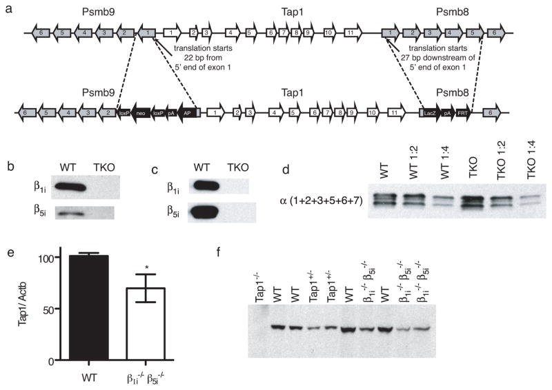
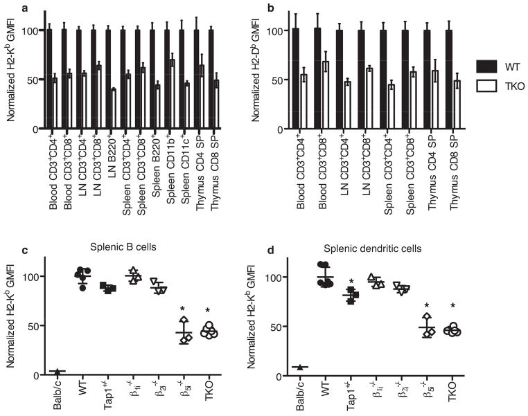
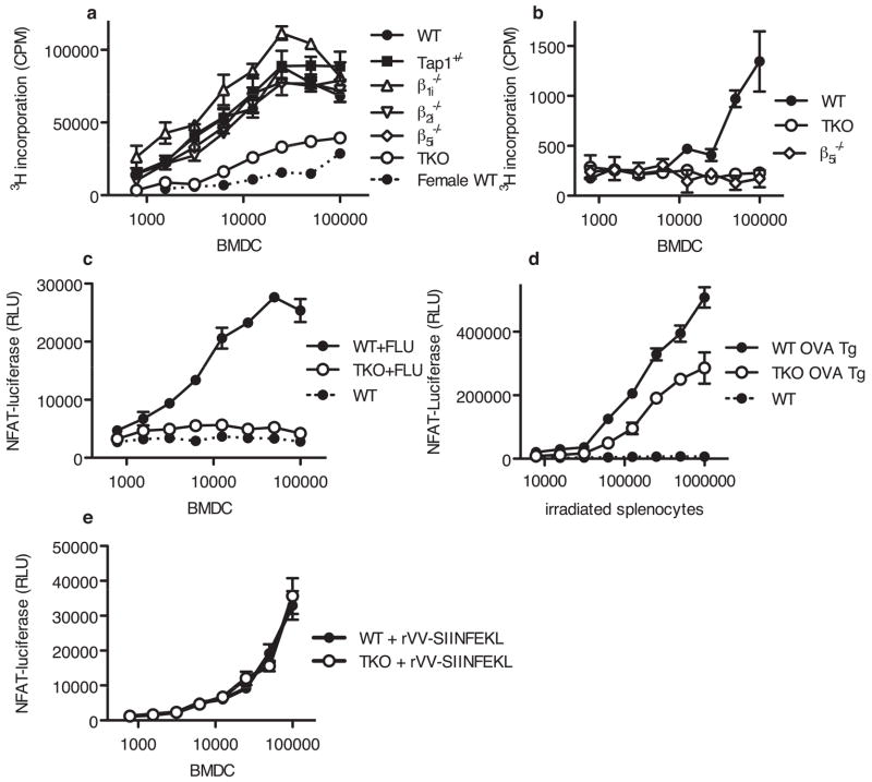
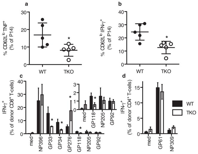
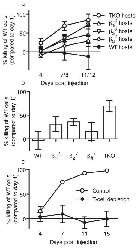
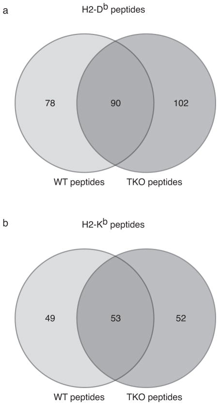
The importance of immunoproteasomes to antigen presentation has been unclear because animals totally lacking immunoproteasomes had not been available. Having now developed mice lacking the three immunoproteasome catalytic subunits, we found that the dendritic cells of these mice had defects in presenting several major histocompatibility complex (MHC) class I epitopes. During viral infection in vivo, the presentation of a majority of MHC class I epitopes was markedly reduced in immunoproteasome-deficient animals compared with wild-type animals, whereas presentation of MHC class II peptides was unaffected. According to mass spectrometry, the repertoire of MHC class I-presented peptides was ∼50% different from that in wild-type mice, and these differences were sufficient to stimulate robust transplant rejection of wild-type cells in mutant mice. These results indicated that immunoproteasomes were more important in antigen presentation than previously thought.


Conflict of interest statement

COMPETING FINANCIAL INTERESTS

The authors declare no competing financial interests.

Figures

Fig. 1
Fig. 1. β1i and β5i double KO mice
(a) Exons targeted for the sequential KO of both Psmb9 and Psmb8 to generate double deficient mice. β1i and β5i proteins are not detected in spleen cell lysates (b) or proteasomes purified from splenocytes (c) (equivalent protein loaded in each lane). (d) The amount of proteasomes in WT and TKO spleens is similar, as measured by alpha subunits in serial dilutions of proteasome pellets from equal numbers of spleens. (e) Normalized TAP1 mRNA expression in WT and β5iβ1i double KO splenocytes, determined by RT-qPCR. Bar indicates mean +/− SD of 5 animals for each strain. Asterisk indicates P < 0.05 (Two-tailed, unpaired t-test).(f) Immunoblotting of splenocyte whole cell lysates from the indicated strains, using anti-TAP1 antibody (equivalent protein loaded in each lane).
Fig. 2
Fig. 2. MHC class I surface expression in TKO and immunoproteasome single KO animals
Blood or organs were harvested from mice of the indicated strains (a) H2-Kb and (b) H2-Db surface expression on cells from WT and TKO mice. In both cases, MHC class I surface expression in TKO mice are significantly reduced compared to WT mice. (Two-way ANOVA, P < 0.0001, n between 3 and 33 depending on the strain and tissue). Bar graphs indicate mean +/− SD. H2-Kb surface expression on splenocytes gated on B220+ (B cells) (c) or CD11c (dendritic cells) (d). Scatter plots indicate individual animals with mean +/− SD indicated. Asterisks indicate a significant difference from WT (P < 0.05, One-way ANOVA followed by Dunnet’s Multiple Comparison Test, n of 3 to 6 for each strain). All results are normalized to the average of WT controls in each experiment.
Fig. 3
Fig. 3. Presentation of H-Y antigens, influenza and OVA by immunoproteasome triple KO dendritic cells is decreased
(a) BMDC from the indicated mouse strains (male unless otherwise indicated) were serially diluted and combined with 3×10⁴ purified HY CD8+ T cells per well. After 3 d of co-incubation, 3H-Thymidine was added and cells were harvested 5 h later. Representative of 4 experiments. (b) Uty antigen presentation in male BMDC of the indicated strains was assayed using 11p9z hybridoma cells. Secreted IL-2 was measured using the CTLL bioassay. Representative of 3 experiments. (c) After a 4 h influenza infection NP366 antigen presentation by infected WT and TKO BMDC was assayed by co-incubating with 12.64-CD8αβ-LUC hybridoma for 12 h. Relative luciferase units (RLU) are shown. Representative of 4 experiments. (d) SIINFEKL antigen presentation by irradiated splenocytes from WT, OVA transgenic or TKO OVA transgenic animals was assayed using RF33.70-LUC hybridoma cells. Representative of 2 experiments. (e) WT and TKO BMDC were infected with rVV-SIINFEKL and fixed after 2 h. SIINFEKL presentation was assayed using RF33.70-LUC hybridoma cells. Representative of 2 experiments. For panels A through D, responses to TKO cells were significantly different from responses to WT cells (Two-way ANOVA, P < 0.0001). Responses were not significantly different between WT and TKO cells in panel E. Error bars indicate S.D. of triplicate values.
Fig. 4
Fig. 4. Altered T cell responses in LCMV-infected TKO mice
(a,b)Two days post LCMV infection, WT and TKO animals were injected with BFA and BFA-treated P14 Tg splenocytes. Spleens were harvested 4 hours later, stained (surface and intracellular) and analyzed by flow cytometry. Representative of 3 experiments. (c,d)To prevent rejection of adoptively transferred cells, host T cells were depleted in WT Thy1.1+Thy1.2+ and TKO Thy1.1+Thy1.2+ mice using anti-Thy1.2. 3×10⁷ LCMV-immune WT Thy1.1 splenocytes were injected i.v. one day before LCMV infection. 5.5 days post-infection, animals were sacrificed and spleens were harvested. Cells analyzed by in vitro restimulation followed by surface and intracellular cytokine staining. Mean and standard deviation of 3 animals of each strain, representative of two experiments. Inset shows low abundance CD8 T-cell responses, enlarged from (c). Asterisk indicated P<0.05, by two-tailed, unpaired t-test.
Fig. 5
Fig. 5. TKO hosts reject WT splenocytes
(a) Mice of the indicated strain were injected with WT and KO cells differentially labeled with CFSE. Mice were bled at the indicated times, and the selective killing of WT cells was assessed. Mean ±SD from: TKO, 14 mice, 3 experiments; β1i, 10 mice, 3 experiments; β5i, 5 mice, 1 experiment; β2i, 7 mice, 2 experiments. (b) Killing of WT splenocytes by single and TKO mice at day 7 or 8 (One way ANOVA, with Dunnett’s Multiple Comparison test, asterisk indicates P < 0.05). (c) Similar to (a) except the TKO host T cells were depleted with anti-Thy1.2 antibody (30H12) before injection of WT splenocytes.
Fig. 6
Fig. 6. Distinct peptides presented on WT and TKO splenocytes
Comparison of peptides identified by tandem mass spectrometry (MS/MS) in WT and TKO animals. pMHCI complexes were isolated with anti H2-Db (a) or anti H2-Kb (b) antibodies. Numbers indicate the number of identified peptides that were unique to WT splenocytes, unique to TKO splenocytes or shared by both.

Comment in

Similar articles

Cited by

References

    1. Rock KL, et al. Inhibitors of the proteasome block the degradation of most cell proteins and the generation of peptides presented on MHC class I molecules. Cell. 1994;78:761–771. - PubMed
    1. Coux O, Tanaka K, Goldberg AL. Structure and functions of the 20S and 26S proteasomes. Annu Rev Biochem. 1996;65:801–847. - PubMed
    1. Tanaka K. Role of proteasomes modified by interferon-gamma in antigen processing. J Leukoc Biol. 1994;56:571–575. - PubMed
    1. Gaczynska M, Rock KL, Goldberg AL. Gamma-interferon and expression of MHC genes regulate peptide hydrolysis by proteasomes. Nature. 1993;365:264–267. - PubMed
    1. Driscoll J, Brown MG, Finley D, Monaco JJ. MHC-linked LMP gene products specifically alter peptidase activities of the proteasome. Nature. 1993;365:262–264. - PubMed

Publication types

Substances