

Is the spinal motion segment a diarthrodial polyaxial joint: what a nice nucleus like you doing in a joint like this?
- PMID: 22197996
- PMCID: PMC3278538
- DOI: 10.1016/j.bone.2011.12.004
Is the spinal motion segment a diarthrodial polyaxial joint: what a nice nucleus like you doing in a joint like this?
Abstract
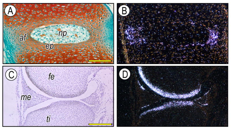
This review challenges an earlier view that the intervertebral joint could not be classified as a diarthrodial joint and should remain as an amphiarthrosis. However, a careful analysis of the relevant literature and in light of more recent studies, it is clear that while some differences exist between the spinal articulation and the generic synovial joint, there are clear structural, functional and developmental similarities between the joints that in sum outweigh the differences. Further, since the intervertebral motion segment displays movement in three dimensions and the whole spine itself provides integrated rotatory movements, it is proposed that it should be classified not as an amphiarthrose, "a slightly moveable joint" but as a complex polyaxial joint. Hopefully, reclassification will encourage further analysis of the structure and function of the two types of overlapping joints and provide common new insights into diseases that afflict the many joints of the human skeleton.
Copyright © 2011 Elsevier Inc. All rights reserved.
Figures

References
-
- Gray H. Anatomy: descriptive and surgical. Philadelphia: Blanchard and Lea; 1859.
-
- Yong-Hing K, Kirkaldy-Willis WH. The pathophysiology of degenerative disease of the lumbar spine. Orthop Clin North Am. 1983;14:491–504. - PubMed
-
- Panjabi MM, Oxland TR, Yamamoto I, Crisco JJ. Mechanical behavior of the human lumbar and lumbosacral spine as shown by three-dimensional load-displacement curves. J Bone Joint Surg Am. 1994;76:413–24. - PubMed
-
- Luschka Hubert. Die Halbgelenke des menschlichen Körpers: Mit 6 Kupfertafeln. Druck Und Verlag Von Georg Reimer; Berlin: 1858.
-
- Grignon B, Roland J. Can the human intervertebral disc be compared to a diarthrodial joint? Surg Radiol Anat. 2000;22:101–5. - PubMed
Publication types
MeSH terms
Grants and funding
LinkOut - more resources
Full Text Sources
