

Role of epithelial mucins during airway infection
- PMID: 22198062
- PMCID: PMC3342466
- DOI: 10.1016/j.pupt.2011.12.003
Role of epithelial mucins during airway infection
Abstract
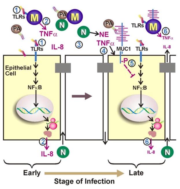
Airway surface fluid contains two layers of mucins consisting mainly of 5 different mucin gene products. While the outer layer contains two gel-forming mucins (MUC5AC and MUC5B) that are tightly associated with various biologically active, defensive molecules, the inner layer contains three membrane-tethered mucins (MUC1, MUC4 and MUC16) shed from the apical cell surface. During airway infection, all of these mucins serve as a major protective barrier against pathogens. MUC1 mucin produced by virtually all the surface columnar epithelial cells in the respiratory tract as well as Type II pneumocytes in the alveoli plays an additional, perhaps more critical role during respiratory infection by controlling the resolution of inflammation that is essential to prevent the development of inflammatory lung disease.
Copyright © 2012 Elsevier Ltd. All rights reserved.
Figures
References
-
- Hollingsworth MA, Swanson BJ. Mucins in cancer: protection and control of the cell surface. Nat Rev Cancer. 2004;4:45–60. - PubMed
-
- Thornton DJ, Rousseau K, McGuckin MA. Structure and function of the polymeric mucins in airways mucus. Annu Rev Physiol. 2008;70:459–86. - PubMed
-
- Rose MC, Voynow JA. Respiratory tract mucin genes and mucin glycoproteins in health and disease. Physiol Rev. 2006;86:245–78. - PubMed
Publication types
MeSH terms
Substances
Grants and funding
LinkOut - more resources
Full Text Sources
Other Literature Sources
Research Materials
Miscellaneous
