. 2012 Feb 1;518(1):71-8.
doi: 10.1016/j.abb.2011.11.024. Epub 2011 Dec 16.

PPARα activation inhibits endothelin-1-induced cardiomyocyte hypertrophy by prevention of NFATc4 binding to GATA-4

Affiliations

PPARα activation inhibits endothelin-1-induced cardiomyocyte hypertrophy by prevention of NFATc4 binding to GATA-4

Kang Le et al. Arch Biochem Biophys. .

Abstract

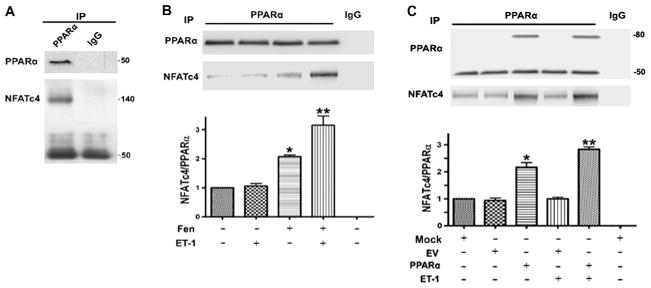
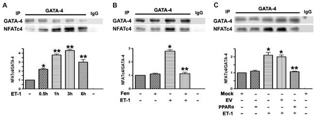
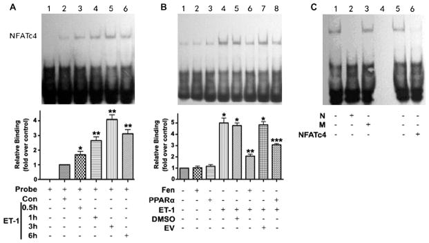
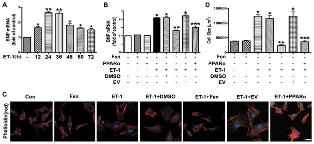
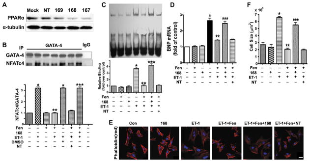
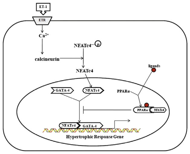
Peroxisome proliferator-activated receptor alpha (PPARα) has been implicated in the pathogenesis of cardiac hypertrophy, although its mechanism of action remains largely unknown. To determine the effect of PPARα activation on endothelin-1 (ET-1)-induced cardiomyocyte hypertrophy and explore its molecular mechanisms, we evaluated the interaction of PPARα with nuclear factor of activated T-cells c4 (NFATc4) in nuclei of cardiomyocytes from neonatal rats in primary culture. In ET-1-stimulated cardiomyocytes, data from electrophoretic mobility-shift assays (EMSA) and co-immunoprecipitation (co-IP) revealed that fenofibrate (Fen), a PPARα activator, in a concentration-dependent manner, enhanced the association of NFATc4 with PPARα and decreased its interaction with GATA-4, in promoter complexes involved in activation of the rat brain natriuretic peptide (rBNP) gene. Effects of PPARα overexpression were similar to those of its activation by Fen. PPARα depletion by small interfering RNA abolished inhibitory effects of Fen on NFATc4 binding to GATA-4 and the rBNP DNA. Quantitative RT-PCR and confocal microscopy confirmed inhibitory effects of PPARα activation on elevation of rBNP mRNA levels and ET-1-induced cardiomyocyte hypertrophy. Our results suggest that activated PPARα can compete with GATA-4 binding to NFATc4, thereby decreasing transactivation of NFATc4, and interfering with ET-1 induced cardiomyocyte hypertrophy.


Figures

Fig. 1
Fig. 1
Activation or overexpression of PPARα enhanced the association of NFATc4 with PPARα in the nuclei of cardiomyocytes. (A) Proteins precipitated from nuclei of rat cardiomyocytes with antibodies against PPARα or control mouse IgG were separated in 8% gels before Western blotting with antibodies against PPARα or NFATc4. Data were similar in two other experiments. (B) Cardiomyocytes were incubated without or with 10 μM fenofibrate (Fen) 1 h and/or 100 nM ET-1 3 h, before IP of nuclear proteins with antibodies against PPARα. Proteins separated by PAGE were quantified by densitometry of Western blots. Data are means ± S.E.M. of values from three independent experiments. *P < 0.01 vs. no additions, **P < 0.01 vs. Fen alone. (C) For overexpression of PPARα-EGFP (80 kDa), cardiomyocytes were incubated 24 h with vehicle (Mock), empty vector (EV), or PPARα-EGFP (PPARα), followed by 3 h without or with 100 nM ET-1 as indicated. Proteins precipitated from nuclei with antibodies against PPARα were separated and analyzed as in Fig. 1B. Data are means ± S.E.M. of values from three independent experiments.*P < 0.001 vs. Mock, **P < 0.01 vs. PPARα.
Fig. 2
Fig. 2
Activation or overexpression of PPARα inhibited the interaction between NFATc4 and GATA-4. (A) Cardiomyocytes were incubated without or with 100 nM ET-1 for indicated times, before IP of nuclear proteins with GATA-4 antibodies or control rabbit IgG. Proteins were separated in 8% gels before Western blotting with antibodies against NFATc4 and GATA-4. Data are means ± S.E.M. of values from three independent experiments. *P < 0.01, **P < 0.001 vs. no additions. (B) Cardiomyocytes were incubated without or with 10 μM Fen (1 h) and/or 100 nM ET-1 (3 h), before IP of nuclear proteins with antibodies against GATA-4. Proteins separated by PAGE were quantified by densitometry of Western blots. Data are means ± S.E.M. of values from three independent experiments. *P < 0.001 vs. no additions, **P < 0.001 vs. ET-1. (C) In studies involving PPARα overexpression, cardiomyocytes were incubated for 24 h with vehicle, empty vector, or PPARα-EGFP, followed by 3 h without or with 100 nM ET-1 as indicated. Proteins precipitated from nuclei with antibodies against GATA-4 were separated and analyzed as in Fig. 2B. Data are means ± S.E.M. of values from three independent experiments. *P < 0.001 vs. Mock, **P < 0.001 vs. ET-1 + EV.
Fig. 3
Fig. 3
The binding activity of NFATc4 to rBNP promoter (−330/−351bp) was decreased by PPARα activation or overexpression in the nuclei of cardiomyocytes. (A) Cardiomyocytes were incubated without or with 100 nM ET-1 for 0.5, 1, 3, or 6 h as indicated before EMSA. Con (control) means without ET-1. The amounts of NFATc4–DNA complexes were quantified by densitometry. Data are means ± S.E.M. of values from three independent experiments. *P < 0.01, **P < 0.001 vs. no additions. (B) EMSA was used to assess binding of NFATc4 with probe in cardiomyocytes treated as described in Fig. 1. The amounts of NFATc4–DNA complexes were quantified by densitometry. Data are means ± S.E.M. of values from three independent experiments. *P < 0.001 vs. no additions, **P < 0.001 vs. ET-1 + DMSO, ***P < 0.001 vs. ET-1 + EV. (C) Effects of unlabeled (cold) probes or NFATc4 antibodies on EMSA. Cold normal (N) or mutant (M) probe was present at 400-fold the concentration of labeled probe, where indicated. Supershift assay were performed by preincubating the nuclear extracts with anti-NFATc4 antibodies (NFATc4). Data were similar in two other experiments.
Fig. 4
Fig. 4
Fen inhibited the co-IP of NFATc4 with GATA-4 and DNA binding to the rBNP promoter in a concentration-dependent manner. Cells were incubated for 1 h with the indicated concentration of Fen, followed by 3 h with 100 nM ET-1 before IP with anti-PPARα antibodies or control IgG, for Western blotting of proteins with antibodies against NFATc4 or PPARα and densitometric quantitation. Data are means ± S.E.M. of values from three independent experiments. *P < 0.001 vs. no additions. (B) Cardiomyocytes were incubated with Fen and/or ET-1 as in Fig. 4A, before IP with antibodies against GATA-4 or control IgG, for Western blotting of proteins with antibodies against NFATc4 or GATA-4, and densitometric quantitation. Data are means ± S.E.M. of values from three independent experiments.*P < 0.001 vs. no additions, **P < 0.001 vs. ET-1. (C) EMSA contained nuclear extracts from cardiomyocytes incubated with Fen and/or ET-1. The amounts of NFATc4-DNA complexes were quantified by densitometry. Data are means ± S.E.M. of values from three independent experiments.*P < 0.001 vs. no additions, **P < 0.001 vs. ET-1.
Fig. 5
Fig. 5
Activation or overexpression of PPARα attenuated ET-1 induced BNP mRNA upregulation and cardiomyocyte hypertrophy. (A) Levels of BNP mRNA in rat cardiomyocytes treated with 100 nM ET-1 for indicated times were assessed by quantitative RT-PCR. Data are means ± S.E.M. of values from three independent experiments.*P < 0.01, **P < 0.001 vs. no additions. (B) Total RNA was extracted from cardiomyocytes for BNP mRNA detection after 24 h of indicated treatment. Data are means ± S.E.M. of values from three independent experiments. *P < 0.001 vs. no additions, **P < 0.001 vs. ET-1 + DMSO, ***P < 0.001 vs. ET-1 + EV. (C) and (D) Cardiomyocytes after 48 h of indicated treatment were stained with rhodamine–phalloidin (C), followed by cell surface area quantitation (D). Scale bar, 20 μm. Data are means ± S.E.M. of values from three independent experiments. *P < 0.001 vs. no additions, **P < 0.001 vs. ET-1 + DMSO, ***P < 0.001 vs. ET-1 + EV.
Fig. 6
Fig. 6
PPARα depletion blocked the inhibitory effects of fenofibrate on ET-1 induced BNP mRNA upregulation and cardiomyocyte hypertrophy. (A) Lysates of cardiomyocytes prepared after incubation for 72 h with vehicle alone (Mock), or with non-target siRNA (NT), or one of three (167, 168, 169) PPARα-directed siRNAs were analysed by Western blotting with α-tubulin as loading control. (B) IP with anti-GATA-4 antibodies or control IgG of the nuclear extracts of cardiomyocytes 72 h after siRNA transfection followed by indicated treatments were immunoblotted with antibodies against NFATc4 and GATA-4, and followed by densitometric quantitation. Data are means ± S.E.M. of values from three independent experiments. *P < 0.001 vs. no additions, **P < 0.001 vs. ET-1, ***P < 0.001 vs. ET-1 + Fen. (C) EMSA was performed with nuclear extracts of cardiomyocytes that had been treated as indicated to assess differences in association of NFATc4 with BNP promoter. The amounts of NFATc4–DNA complexes were quantified by densitometry. Data are means ± S.E.M. of values from three independent experiments. *P < 0.001 vs. no additions, **P < 0.001 vs. ET-1, ***P < 0.001 vs. ET-1 + Fen. (D) Total RNA from cardiomyocytes was template for quantitation of BNP mRNA 72 h after siRNA transfection followed by indicated treatments. Data are means ± S.E.M. of values from three independent experiments. *P < 0.001 vs. no additions, **P < 0.001 vs. ET-1, ***P < 0.001 vs. ET-1 + Fen. (E) and (F) Cardiomyocytes 72 h after siRNA transfection following indicated treatments were stained with rhodamine–phalloidin (E), followed by surface area quantitation (F). Scale bar, 20 μm. Data are means ± S.E.M. of values from three independent experiments.*P < 0.001 vs. no additions, **P < 0.001 vs. ET-1, ***P < 0.001 vs. ET-1 + Fen.
Fig. 7
Fig. 7
A model for the function of PPARα activation on cardiomyocyte hypertrophy induced by ET-1. Activated PPARα associates with activated NFATc4 induced by ET-1 in the nucleus to prevent the interaction of NFATc4 with GATA-4, and further reduce the binding of NFATc4 to the promoter of hypertrophic response genes, and thereby inhibit cardiomyocyte hypertrophy.

Similar articles

Cited by

References

    1. Lloyd-Jones DM, Larson MG, Leip EP, Beiser A, D’Agostino RB, Kannel WB, Murabito JM, Vasan RS, Benjamin EJ, Levy D. Circulation. 2002;106:3068–3072. - PubMed
    1. Ho KK, Pinsky JL, Kannel WB, Levy D. Journal of the American College of Cardiology. 1993;22:6A–13A. - PubMed
    1. Finckenberg P, Mervaala E. Journal of Hypertension. 2010;28(Suppl 1):S33–38. - PubMed
    1. Eder P, Molkentin JD. Circulation Research. 2011;108:265–272. - PubMed
    1. Chien KR, Knowlton KU, Zhu H, Chien S. FASEB Journal. 1991;5:3037–3046. - PubMed

Publication types

MeSH terms

LinkOut - more resources