. 2011 Dec 25;481(7382):502-5.
doi: 10.1038/nature10732.

Purkinje neuron synchrony elicits time-locked spiking in the cerebellar nuclei

Affiliations

Purkinje neuron synchrony elicits time-locked spiking in the cerebellar nuclei

Abigail L Person et al. Nature. .

Abstract

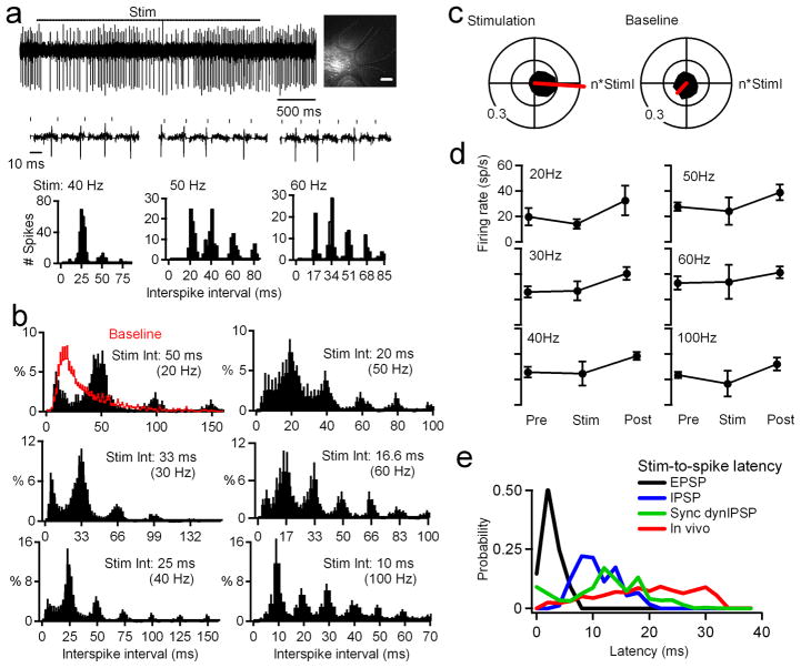
An unusual feature of the cerebellar cortex is that its output neurons, Purkinje cells, release GABA (γ-aminobutyric acid). Their high intrinsic firing rates (50 Hz) and extensive convergence predict that their target neurons in the cerebellar nuclei would be largely inhibited unless Purkinje cells pause their spiking, yet Purkinje and nuclear neuron firing rates do not always vary inversely. One indication of how these synapses transmit information is that populations of Purkinje neurons synchronize their spikes during cerebellar behaviours. If nuclear neurons respond to Purkinje synchrony, they may encode signals from subsets of inhibitory inputs. Here we show in weanling and adult mice that nuclear neurons transmit the timing of synchronous Purkinje afferent spikes, owing to modest Purkinje-to-nuclear convergence ratios (∼40:1), fast inhibitory postsynaptic current kinetics (τ(decay) = 2.5 ms) and high intrinsic firing rates (∼90 Hz). In vitro, dynamically clamped asynchronous inhibitory postsynaptic potentials mimicking Purkinje afferents suppress nuclear cell spiking, whereas synchronous inhibitory postsynaptic potentials entrain nuclear cell spiking. With partial synchrony, nuclear neurons time-lock their spikes to the synchronous subpopulation of inputs, even when only 2 out of 40 afferents synchronize. In vivo, nuclear neurons reliably phase-lock to regular trains of molecular layer stimulation. Thus, cerebellar nuclear neurons can preferentially relay the spike timing of synchronized Purkinje cells to downstream premotor areas.


Figures

Figure 1
Figure 1. Purkinje cell convergence and fast IPSCs in the cerebellar nuclei
a, IPSCs evoked by maximal stimulation. Holding potential −57 mV (junction potential corrected). Top: 5 overlaid IPSCs. Middle: IPSC amplitudes as stimulus strength was reduced. Bottom: Maximal conductances calculated from measured IPSC reversal potentials (n=17, mean ECl=−75 mV). Bin width, 10 nS. b, IPSCs evoked by minimal stimulation (range 16–20 μA). Top: 20 overlaid IPSCs. Middle: Expansion of box in (a). Events near the zero line fell within the noise. Bottom: Minimal conductances (n=30). Bin width, 2 nS. c, Left: Scaled IPSCs in one cell at 22° and 36oC. Right: Temperature dependence of IPSC decay τ (n=7). d, Top: 50-Hz train of IPSCs at 36oC. Bottom: Depression of phasic IPSCs and absence of tonic current during train. In all figures, data are mean±s.e.m, unless noted.
Figure 2
Figure 2. Entrainment of nuclear neuron spiking to synchronous IPSPs
a, Top: Response to desynchronized and synchronized dynIPSPs from 8 inputs at 50 Hz, τdecay =2.5 ms. Bottom: Instantaneous firing rates of neuron in (a), showing entrainment during synchrony. Arrows, onset of desynchronized (left) and synchronized (right) dynIPSPs. Tall spikes reflect overlapping dynIPSPs and spikes. b, Firing rates during desynchronized and synchronized 50-Hz trains with different dynIPSP kinetics (τdecay=2.5, 5, 14 ms, n=25, 18, 10). c, Entrainment to corticonuclear IPSPs (36°C) between 60 and 80 Hz. Arrows, stimulation times. d, Median firing rates (±1 quartile) in response to evoked IPSPs (closed symbols) and desynchronized dynIPSPs (open symbols). From 50–100 Hz, evoked, n=8, 8, 8, 7, 3; simulated, n=25, 10, 10, 9, 14. Dashed line, unity.
Figure 3
Figure 3. The synchronous subpopulation of Purkinje inputs sets spike timing of nuclear neurons
a, Nuclear neuron responses to dynIPSPs from 40 asynchronous or partially synchronized inputs, as labeled. b, Spike rasters during dynIPSP trains with 50–5% synchrony. c, Firing rates during 20–125 Hz trains with 0%–50% synchrony (“sync rates”). d, Normalized interspike interval distributions during 50% synchrony, from 40 to 100 Hz. Abscissa tick marks indicate multiples of the interstimulus intervals. Bin width, 2 ms. Black: no current injection. Blue: with 200 pA DC applied to increase spike number during inhibition. e, Black: Polar histograms of interspike intervals during 50–5% synchrony for 50, 55, 67, 83, 100, and 125 Hz input. Each cycle is one interstimulus interval (StimI). Red: Net vectors of interspike interval histograms. (For 50%, 25%, 10%, 5% synchrony, n=14, 9, 7, 7).
Figure 4
Figure 4. Nuclear neurons phase lock to molecular layer stimulation in vivo
a, Upper trace, response of a nuclear neuron to 40-Hz molecular layer stimulation (bar). Inset, Recording site in the cerebellar nuclei recovered after focal Alexa 568 injection. Dashed lines demarcate cerebellar folia. Scale bar, 200 μm. Lower traces, expanded segments of spikes during 40, 50, and 60-Hz stimulation (ticks), showing the long stimulus-to-spike latency. Stimulus artifacts are zeroed. Plots, Interspike interval histograms for responses above. b, Mean normalized interspike interval distributions during molecular layer stimulation from 20–100 Hz for 4, 9, 9, 7, 5, and 7 cells respectively. Abscissa tick marks indicate multiples of the interstimulus interval. Red baseline histogram includes intervals before and after stimulation. Peaks at multiples of 8.3 ms were evident in 2/2 cells stimulated at 120 Hz (not shown). Bin width, 2 ms. c, Black: Polar histograms of interspike intervals during stimulation across rates (left) or during baseline periods. Red: Net vectors of polar histograms. d, Mean firing rates before (“pre,” 1 sec), during (“stim,” 3 sec) and after (“post,” 9 sec) stimulation. e, Stimulus-to-spike latencies for pharmacologically isolated EPSPs (black), IPSPs (blue), dynIPSPs during partial synchrony (green) and in vivo (red).

Comment in

Similar articles

Cited by

References

    1. Thach WT. Discharge of Purkinje and cerebellar nuclear neurons during rapidly alternating arm movements in the monkey. J Neurophysiol. 1968;31:785–97. - PubMed
    1. Chan-Palay V. Organization, cytology, and transmitters. Springer; Berlin: 1977. Cerebellar dentate nucleus.
    1. Palkovits M, Mezeky E, Hamori J, Szentagothai J. Quantitative histological analysis of the cerebellar nuclei in the cat. I. Numerical data on cells and on synapses. Exp Brain Res. 1977;28:189–209. - PubMed
    1. McDevitt CJ, Ebner TJ, Bloedel JR. Relationships between simultaneously recorded Purkinje cells and nuclear neurons. Brain Res. 1987;425:1–13. - PubMed
    1. Bell CC, Grimm RJ. Discharge properties of Purkinje cells recorded on single and double microelectrodes. J Neurophysiol. 1969;32(6):1044–1055. - PubMed

Publication types