

Spasmolytic polypeptide-expressing metaplasia (SPEM) in the gastric oxyntic mucosa does not arise from Lgr5-expressing cells
- PMID: 22198711
- PMCID: PMC3767919
- DOI: 10.1136/gutjnl-2011-301193
Spasmolytic polypeptide-expressing metaplasia (SPEM) in the gastric oxyntic mucosa does not arise from Lgr5-expressing cells
Abstract
Objective: Metaplastic lineages in the oxyntic mucosa of the stomach are critical preneoplastic precursors of gastric cancer. Recent studies have demonstrated that spasmolytic polypeptide-expressing metaplasia (SPEM) in the mouse oxyntic mucosa arises from transdifferentiation of mature gastric chief cells. Other investigations of intestinal progenitor cells have shown that cells demonstrating transcriptional activity for leucine-rich repeat containing G-protein-coupled receptor 5 (Lgr5) in the intestine, colon and gastric antrum function as adult stem cells. We have now investigated whether cells demonstrating Lgr5 transcriptional activity in the oxyntic mucosa of mice might be responsible for development of metaplasia.
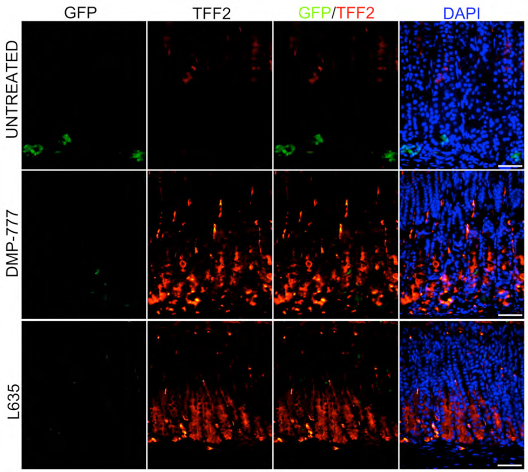
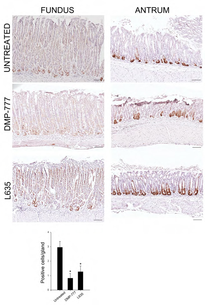
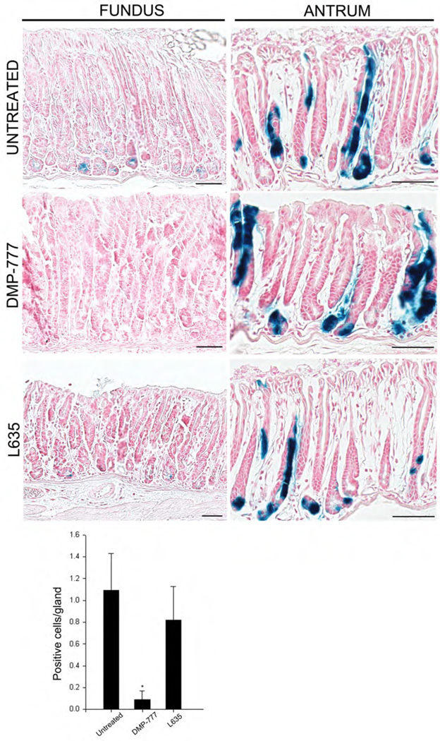
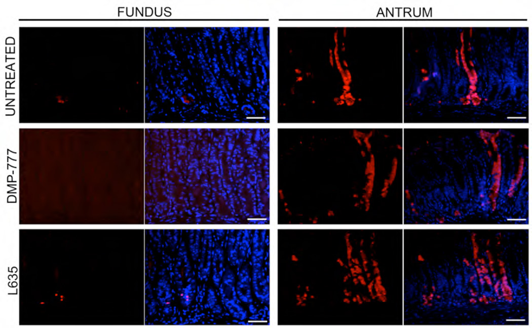
Design: Lgr5-EGFP-IRES-Cre(ERT2/+);Rosa26R mice were used to examine the distribution of Lgr5 transcriptionally active cells in the normal oxyntic mucosa as well as after treatment with DMP-777 or L-635 to induce acute SPEM. Lineage mapping was performed to determine if Lgr5-expressing cells gave rise to SPEM.
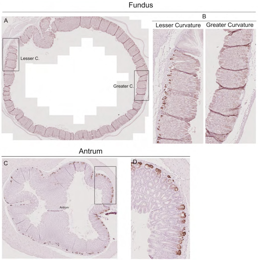
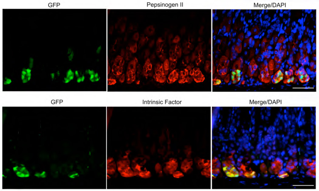
Results: Cells expressing transcriptional activity for Lgr5 in the oxyntic mucosa were present as scattered rare cells only along the lesser curvature of the stomach. These cells also stained for markers of chief cells (intrinsic factor and pepsinogen) but never showed any staining for proliferative markers (Ki-67). In Lgr5-EGFP-IRES-Cre(ERT2/+);Rosa26R mice induced with tamoxifen, treatment with either DMP-777 or L-635 to induce acute oxyntic atrophy caused induction of SPEM, but no lineage mapping into SPEM from Lgr5-expressing cells was observed.
Conclusion: The results indicate that, while chief cells with Lgr5 transcriptional activity are present along the lesser curvature of the gastric oxyntic mucosa, they are not responsible for production of metaplasia.
Conflict of interest statement
None of the authors have any conflicts of interest in the pursuit of this work.
Figures
Comment in
-
Lgr5 expression is absent in human premalignant lesions of the stomach.Gut. 2012 Dec;61(12):1777-8. doi: 10.1136/gutjnl-2012-302372. Epub 2012 Mar 22. Gut. 2012. PMID: 22442165 No abstract available.
Similar articles
-
A molecular signature of gastric metaplasia arising in response to acute parietal cell loss.Gastroenterology. 2008 Feb;134(2):511-22. doi: 10.1053/j.gastro.2007.11.058. Epub 2007 Dec 4. Gastroenterology. 2008. PMID: 18242217 Free PMC article.
-
Potentiation of oxyntic atrophy-induced gastric metaplasia in amphiregulin-deficient mice.Gastroenterology. 2007 May;132(5):1804-19. doi: 10.1053/j.gastro.2007.03.040. Epub 2007 Mar 24. Gastroenterology. 2007. PMID: 17484876
-
Mature gastric chief cells are not required for the development of metaplasia.Am J Physiol Gastrointest Liver Physiol. 2018 May 1;314(5):G583-G596. doi: 10.1152/ajpgi.00351.2017. Epub 2018 Jan 18. Am J Physiol Gastrointest Liver Physiol. 2018. PMID: 29345968 Free PMC article.
-
Differentiation of the gastric mucosa III. Animal models of oxyntic atrophy and metaplasia.Am J Physiol Gastrointest Liver Physiol. 2006 Dec;291(6):G999-1004. doi: 10.1152/ajpgi.00187.2006. Am J Physiol Gastrointest Liver Physiol. 2006. PMID: 17090722 Review.
-
Current understanding of SPEM and its standing in the preneoplastic process.Gastric Cancer. 2009;12(4):189-97. doi: 10.1007/s10120-009-0527-6. Epub 2010 Jan 5. Gastric Cancer. 2009. PMID: 20047123 Free PMC article. Review.
Cited by
-
Intestinal Stem Cell Markers in the Intestinal Metaplasia of Stomach and Barrett's Esophagus.PLoS One. 2015 May 21;10(5):e0127300. doi: 10.1371/journal.pone.0127300. eCollection 2015. PLoS One. 2015. PMID: 25996368 Free PMC article.
-
Regulation of Gastric Lgr5+ve Cell Homeostasis by Bone Morphogenetic Protein (BMP) Signaling and Inflammatory Stimuli.Cell Mol Gastroenterol Hepatol. 2018 Jan 31;5(4):523-538. doi: 10.1016/j.jcmgh.2018.01.007. eCollection 2018. Cell Mol Gastroenterol Hepatol. 2018. PMID: 29930977 Free PMC article.
-
Role and research progress of spasmolytic polypeptide‑expressing metaplasia in gastric cancer (Review).Int J Oncol. 2024 Mar;64(3):33. doi: 10.3892/ijo.2024.5621. Epub 2024 Feb 1. Int J Oncol. 2024. PMID: 38299264 Free PMC article.
-
Distribution of LGR5+ cells and associated implications during the early stage of gastric tumorigenesis.PLoS One. 2013 Dec 10;8(12):e82390. doi: 10.1371/journal.pone.0082390. eCollection 2013. PLoS One. 2013. PMID: 24340024 Free PMC article. Clinical Trial.
-
Dysregulated Immune Responses by ASK1 Deficiency Alter Epithelial Progenitor Cell Fate and Accelerate Metaplasia Development during H. pylori Infection.Microorganisms. 2020 Dec 14;8(12):1995. doi: 10.3390/microorganisms8121995. Microorganisms. 2020. PMID: 33542169 Free PMC article.
References
-
- Karam SM, Leblond CP. Dynamics of epithelial cells in the corpus of the mouse stomach. III. Inward migration of neck cells followed by progressive transformation into zymogenic cells. Anat Rec. 1993;236:297–313. - PubMed
-
- Karam SM. Dynamics of epithelial cells in the corpus of the mouse stomach. IV. Bidirectional migration of parietal cells ending in their gradual degeneration and loss. Anat Rec. 1993;236:314–332. - PubMed
-
- Ramsey VG, Doherty JM, Chen CC, et al. The maturation of mucus-secreting gastric epithelial progenitors into digestive-enzyme secreting zymogenic cells requires Mist1. Development. 2007;134:211–222. - PubMed
-
- Karam SM, Leblond CP. Dynamics of epithelial cells in the corpus of the mouse stomach. I. Identification of proliferative cell types and pinpointing of the stem cells. AnatRec. 1993;236:259–279. - PubMed
-
- Barker N, Huch M, Kujala P, et al. Lgr5(+ve) stem cells drive self-renewal in the stomach and build long-lived gastric units in vitro. Cell Stem Cell. 2010;6:25–36. - PubMed
Publication types
MeSH terms
Substances
Grants and funding
LinkOut - more resources
Full Text Sources
Medical