. 2012 Jan 3;109(1):119-24.
doi: 10.1073/pnas.1114089108. Epub 2011 Dec 23.

Stabilized G protein binding site in the structure of constitutively active metarhodopsin-II

Affiliations

Stabilized G protein binding site in the structure of constitutively active metarhodopsin-II

Xavier Deupi et al. Proc Natl Acad Sci U S A. .

Abstract

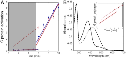
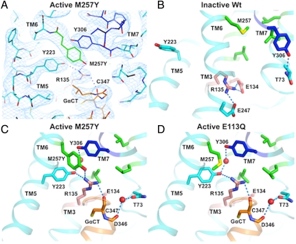
G protein-coupled receptors (GPCR) are seven transmembrane helix proteins that couple binding of extracellular ligands to conformational changes and activation of intracellular G proteins, GPCR kinases, and arrestins. Constitutively active mutants are ubiquitously found among GPCRs and increase the inherent basal activity of the receptor, which often correlates with a pathological outcome. Here, we have used the M257Y(6.40) constitutively active mutant of the photoreceptor rhodopsin in combination with the specific binding of a C-terminal fragment from the G protein alpha subunit (GαCT) to trap a light activated state for crystallization. The structure of the M257Y/GαCT complex contains the agonist all-trans-retinal covalently bound to the native binding pocket and resembles the G protein binding metarhodopsin-II conformation obtained by the natural activation mechanism; i.e., illumination of the prebound chromophore 11-cis-retinal. The structure further suggests a molecular basis for the constitutive activity of 6.40 substitutions and the strong effect of the introduced tyrosine based on specific interactions with Y223(5.58) in helix 5, Y306(7.53) of the NPxxY motif and R135(3.50) of the E(D)RY motif, highly conserved residues of the G protein binding site.


Conflict of interest statement

The authors declare no conflict of interest.

Figures

Fig. 1.
Fig. 1.
Biochemical assessment of M257Y6.40 in the context of the N2C/D282C background. (A) Assessment of G protein activation (pmol) by bovine opsin (open symbols and dashed regression lines) or 11-cis-retinal reconstituted rhodopsin (solid symbols and regression lines) for the N2C/D282C (blue circles) and N2C/M257Y/D282C (red triangles) mutants. The shaded area represents dim red light conditions that preserve the inactive ground state of rhodopsin. At time 5 min 30 s, samples were subjected to bright illumination and the rest of the data were collected. (B) Ability of opsin to covalently bind all-trans-retinal. Absorbance spectra of purified N2C/M257Y/D282C reconstituted with all-trans-retinal were recorded at pH 7.4 (solid line) and pH 3.5 (dotted line). At pH 7.4, the pigment absorbs maximally at 380 nm, which is characteristic of metarhodopsin-II and free retinal. Acidification causes a red-shift to 415 nm, resulting from the protonation of the Schiff base formed between opsin and retinal. The inset shows that reconstitution of N2C/M257Y/D282C with all-trans-retinal (squares, dotted regression line, slope = 1.37 pmol/ min) increases G protein activation levels to those of fully light activated metarhodopsin-II that had been reconstituted with 11-cis-retinal (red triangles, solid regression line, slope = 1.35 pmol/ min). Error bars in (A) and (B) represent standard error, n = 3.
Fig. 2.
Fig. 2.
Effect of the M257Y6.40 mutation on the ionic lock region. The 2Fo-Fc density map (A, blue mesh, contoured at 1.5σ) indicates a high degree of order around the NPxxY (dark blue) and E(D)RY motifs (salmon) that are close to the binding site for the G protein peptide GαCT (orange). The bulky side chains of Y2235.58 and Y3067.53, as well as the mutated side chain of Y2576.40 can be accurately positioned within their respective densities. In the ground state of wild-type rhodopsin (B), M2576.40 is part of the hydrophobic barrier (green) that separates the NPxxY motif from the ionic lock region including the E(D)RY motif. The M257Y6.40 mutation (C) interferes with this packing and lowers the energy barrier for reorganizations of TM6 that characterize the active conformation. In the structure of the constitutively active E113Q3.28 structure (D) the hydrophobic barrier has opened and Y2235.58 in TM5 and Y3067.53 of the NPxxY motif in TM7 swing into the created cavity, stabilized by a water-mediated H-bond network (9). In both active state structures (C + D), the ionic lock interactions between R1353.50 and E1343.49 of the E(D)RY motif in TM3 and E2476.30 in TM6 are opened allowing R1353.50 to adopt an elongated rotamer that forms part of the GαCT peptide binding site. The constitutively active M257Y6.40 mutant (C) introduces only minor changes to the overall structure of the ionic lock region, however the introduced tyrosine forms an edge-face interaction with Y2235.58, a parallel-displaced π-π stacking interaction to Y3067.53, and hydrophilic interactions to R1353.50, three interactions that stabilize the open G protein binding site.
Fig. 3.
Fig. 3.
Conformation of all-trans-retinal in the M257Y/GαCT structure. The 2Fo-Fc density map contoured at 1σ (A, blue mesh) shows clear uninterrupted density for all-trans-retinal including the covalent bond to K2967.43 that is characteristic for the active metarhodopsin-II state. The retinal binding pocket (B, C, defined as residues with interatomic distances to the retinal ≤ 4 ) is dominated by hydrophobic van der Waals contacts between the β-ionone ring and four amino acids in TM5 (green), three amino acids in TM6 (blue), and one in EL2 (violet). The polyene part of the retinal is covalently bound to K2967.43 in TM7 (red) and stacked against Y2686.51 in TM6 (blue), while the two amino acids in TM3 (orange) directly interact with the C19 and C20 methyl groups.

References

    1. Ye S, et al. Tracking G-protein-coupled receptor activation using genetically encoded infrared probes. Nature. 2010;464:1386–1389. - PubMed
    1. Palczewski K, et al. Crystal structure of rhodopsin: A G protein-coupled receptor. Science. 2000;289:739–745. - PubMed
    1. Li J, Edwards PC, Burghammer M, Villa C, Schertler GF. Structure of bovine rhodopsin in a trigonal crystal form. J Mol Biol. 2004;343:1409–1438. - PubMed
    1. Standfuss J, et al. Crystal structure of a thermally stable rhodopsin mutant. J Mol Biol. 2007;372:1179–1188. - PMC - PubMed
    1. Nakamichi H, Okada T. Crystallographic analysis of primary visual photochemistry. Angew Chem Int Ed Engl. 2006;45:4270–4273. - PubMed

Publication types

Associated data

LinkOut - more resources