. 2012 Mar 15;302(6):L541-54.
doi: 10.1152/ajplung.00282.2011. Epub 2011 Dec 23.

Loss-of-function thrombospondin-1 mutations in familial pulmonary hypertension

Affiliations

Loss-of-function thrombospondin-1 mutations in familial pulmonary hypertension

James P Maloney et al. Am J Physiol Lung Cell Mol Physiol. .

Abstract

Most patients with familial pulmonary arterial hypertension (FPAH) carry mutations in the bone morphogenic protein receptor 2 gene (BMPR2). Yet carriers have only a 20% risk of disease, suggesting that other factors influence penetrance. Thrombospondin-1 (TSP1) regulates activation of TGF-β and inhibits endothelial and smooth muscle cell proliferation, pathways coincidentally altered in pulmonary arterial hypertension (PAH). To determine whether a subset of FPAH patients also have mutations in the TSP1 gene (THBS1) we resequenced the type I repeats of THBS1 encoding the TGF-β regulation and cell growth inhibition domains in 60 FPAH probands, 70 nonfamilial PAH subjects, and in large control groups. We identified THBS1 mutations in three families: a novel missense mutation in two (Asp362Asn), and an intronic mutation in a third (IVS8+255 G/A). Neither mutation was detected in population controls. Mutant 362Asn TSP1 had less than half of the ability of wild-type TSP1 to activate TGF-β. Mutant 362Asn TSP1 also lost the ability to inhibit growth of pulmonary arterial smooth muscle cells and was over threefold less effective at inhibiting endothelial cell growth. The IVS8+255 G/A mutation decreased and/or eliminated local binding of the transcription factors SP1 and MAZ but did not affect RNA splicing. These novel mutations implicate THBS1 as a modifier gene in FPAH. These THBS1 mutations have implications in the genetic evaluation of FPAH patients. However, since FPAH is rare, these data are most relevant as evidence for the importance of TSP1 in pulmonary vascular homeostasis. Further examination of THBS1 in the pathogenesis of PAH is warranted.


Figures
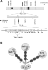
Fig. 1.
Fig. 1.
A: THBS1 gene, including chromosomal neighbors. All exons (rectangles) and introns (lines) are depicted. Exons 7–9 (type I repeats) were resequenced for this project. Direction of gene translation is per arrows. B: trimeric THBS1 protein (TSP1). N, globular region N (NH2-terminal); C, procollagen homology domain; TSR1, 2, and 3, thrombospondin type I repeats; E1, 2, 3, EGF homology (type II) repeats; CaG, calmodulin homology (type III) repeats; G, globular domain (COOH-terminal). The location of the missense mutation found in TSR1 is designated with an asterisk (*). Exon sizes are relative to each other but are exaggerated for display.
Fig. 2.
Fig. 2.
THBS1 mutations found in familial pulmonary arterial hypertension (FPAH) subjects. A: mutation location, type, and chromatograms. B: confirmation of the Asp362Asn mutation by restriction fragment length polymorphism analysis (RFLP). A BccI site is present in the wild-type allele, cutting PCR products in those subjects into fragments of 62 and 223 base pairs (bp) (lanes 14). Only a single strand cuts in mutation carriers (lanes 56; arrow denotes uncut 285-bp fragment). C: Clustal alignment in the region of the Asp362Asn mutation in TSR1. Letters are standard amino acid abbreviations. Left is NH2-terminal. Asp362 (arrow) and flanking residues are highly conserved in vertebrates. Two overlapping WXXW domains are near Asp362.
Fig. 3.
Fig. 3.
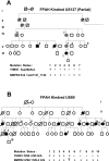
Transmission of THBS1 mutations and associated BMPR2 mutations in FPAH cohorts. A: US127 family with known exon 9 BMPR2 mutation. Only a partial family tree is depicted. The parental pair on the right had 7 offspring, indicated by the dashed line; 12 descendants had PAH associated with the BMPR2 mutation, but the 7 who donated specimens were wild-type for THBS1. B: US80 family with known IVS8 BMPR2 mutation. Symbol key: 1, proband; circle, female; square, male; solid symbols, PAH diagnosis; slash through symbol, deceased; circle within symbol (target), obligate carrier of BMPR2 mutation. Generations are in Roman numerals where space allows.
Fig. 4.
Fig. 4.
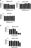
The 362Asn TSP1 mutation displays loss-of-function characteristics on latent TGF-β1 activation in the mink lung epithelial cell bioassay. A: latent TGF-β1 activation by synthetic 13-amino acid TSR1 peptides centered on the 362Asn TSP1 locus. At all concentrations wild-type TSR1 (362Asp) is a more potent activator compared with mutant TSR1 [362Asn, which activates more than scrambled peptide (SCR) only when ≥5 nM]. 362Asp effects were always greater than SCR effects (*P < 0.05). B: trimeric recombinant TSP1 (rTSP1) is a more potent activator of TGF-β1 (pM effects). The mutant isoform is weaker at all concentrations compared with the wild-type isoform (*P < 0.01, **P < 0.001). Activation is expressed as fold change compared with vehicle (zero). Symbols indicate statistical significance by ANOVA or unpaired t-tests. Results represent summed means ± SD of ≥4 experiments.
Fig. 5.
Fig. 5.
THBS1 Asp362Asn mutation effects on inhibition of endothelial cell (HPAEC) proliferation at 72 h. A: effects of 13-amino acid TSR1 peptides. Wild-type 362Asp peptides inhibited proliferation at concentrations ≥0.5 nM; although mutant 362Asn and SCR peptides had no effect. B: effects of rTSP1 on proliferation. Wild-type rTSP1 had more potent effects than mutant rTSP1. Results expressed as means ± SD, summed results of 4 experiments. *P < 0.05 **P < 0.01 vs. vehicle effects.
Fig. 6.
Fig. 6.
362Asn mutation displays loss-of-function properties on TSP1 inhibition of human pulmonary artery smooth muscle cell (HPASMC) proliferation at 72 h. Full-length rTSP1 was used for all studies. Only wild-type 362Asp rTSP1 had significant inhibitory effects. Stimulatory effects on proliferation were seen with both isoforms at low concentrations. Results are expressed as means ± SD, summed results of 4 experiments. *P < 0.05 **P < 0.01 vs. vehicle (zero) values.
Fig. 7.
Fig. 7.
EMSAs evaluating binding of proteins from vascular cell nuclear extracts (NE) and recombinant SP1 and MAZ proteins (rSP1, rMAZ) to biotin-labeled oligonucleotides (oligos) of the THBS1 IVS8+255A mutation region. Labeled oligos are depicted at the top of figures, including positive controls. NE are from HPASMC (SMC) or HPAEC (Endo). Unlabeled competitors were added at 100-fold excess to assess specificity or competition. A: mutant IVS8-A oligos (MUT; lanes 68) bind Endo-NE less than wild-type oligos (WT; lanes 13). Excess unlabeled IVS8-A oligos remain an effective inhibitor of labeled-IVS8-G oligo binding to Endo-NE, consistent with some protein binding being maintained by the mutant sequence (lane 5). B: IVS8 oligo binding to SMC-NE was not different (P = 0.39). Excess of either unlabeled oligo abrogates binding (lanes 5 and 10). C: IVS8-A oligos bind rSP1 poorly (lanes 810). rSP1 binds avidly to a positive control SP1 oligo (lanes 13) and to IVS8-G oligos (lanes 46) in a specific manner (lane 7). D: IVS8-A oligos bind rMAZ poorly (lanes 8 and 9) compared with avid binding to IVS8-G oligos (lanes 46; specificity shown in lane 7) and to rMAZ positive control oligos (lanes 13). Arrows indicate shifted bands; brackets indicate free labeled probe. Analysis of band densities from all experiments is depicted by graphs to the right. Results are expressed as means ± SD as the sum of 3 experiments. Radiographs are representative. *P < 0.05; **P < 0.01, or ***P < 0.001 vs. baseline values. The presence of 2 shifted bands with rMAZ and rSP1 (vs. 1 band with NE) in EMSA is related to the tendency of these purified proteins to form both monomers and dimers.
Fig. 8.
Fig. 8.
A: data from chromatin immunoprecipitation (ChIP) experiments using chromatin from normal HPASMC grown under standard conditions. Chromatin was fixed and sheared, and then specific antibodies to SP1 or MAZ were used to pull down chromatin that endogenously bound SP1 or MAZ proteins (rabbit IgG was control antibody). Immunoprecipitated chromatin was amplified with primers flanking the THBS1 IVS8+255 mutation; then equal aliquots of PCR products were run on an agarose gel, shown at the top of the representative figure (ethidium-stained gel; empty, empty lane; input, amplification of input chromatin, never treated with antibody; DNA bp size marker is in leftmost lane). The fold enrichment of immunoprecipitated chromatin (relative to control IgG antibody) for specific proteins is shown in graph at bottom as determined by quantitative SYBR-green PCR (sum of 6 experiments, means ± SD; * P < 0.05, ** P < 0.01). B: analysis of THBS1 IVS8+255 G/A site occupancy in mutant IVS8+255A carrier lymphoblasts after ChIP as in A. After PCR amplification of the THBS1 IVS8 region, PCR products were subcloned and 10 randomly selected colonies (single clones) were isolated and sequenced for IVS8+255 genotype. Input DNA contained both alleles, whereas immunoprecipitation for SP1 or MAZ revealed only the presence of the G allele, demonstrating that that the A allele disrupted binding of SP1 and MAZ to THBS1 DNA at this locus in intact cells (P < 0.006; 2-tailed χ2).

Similar articles

Cited by

References

    1. Abe M, Harpel JG, Metz CN, Nunes I, Loskutoff DJ, Rifkin DB. An assay for transforming growth factor-beta using cells transfected with a plasminogen activator inhibitor-1 promoter-luciferase construct. Anal Biochem 216: 276–284, 1994 - PubMed
    1. Almeida-Vega S, Catlow K, Kenny S, Dimaline R, Varro A. Gastrin activates paracrine networks leading to induction of PAI-2 via MAZ and ASC-1. Am J Physiol Gastrointest Liver Physiol 296: G414–G423, 2009 - PMC - PubMed
    1. Ashfield R, Patel AJ, Bossone SA, Brown H, Campbell RD, Marcu KB, Proudfoot NJ. MAZ-dependent termination between closely spaced human complement genes. EMBO J 13: 5656–5667, 1994 - PMC - PubMed
    1. Bossone SA, Asselin C, Patel AJ, Marcu KB. MAZ, a zinc finger protein, binds to c-MYC and C2 gene sequences regulating transcriptional initiation and termination. Proc Natl Acad Sci USA 89: 7452–7456, 1992 - PMC - PubMed
    1. Botella LM, Sanchez-Elsner T, Rius C, Corbi A, Bernabeu C. Identification of a critical Sp1 site within the endoglin promoter and its involvement in the transforming growth factor-beta stimulation. J Biol Chem 276: 34486–34494, 2001 - PubMed

Publication types

MeSH terms

Substances