. 2012 Feb 1;188(3):1503-13.
doi: 10.4049/jimmunol.1102832. Epub 2011 Dec 23.

IL-33-responsive lineage- CD25+ CD44(hi) lymphoid cells mediate innate type 2 immunity and allergic inflammation in the lungs

Affiliations

IL-33-responsive lineage- CD25+ CD44(hi) lymphoid cells mediate innate type 2 immunity and allergic inflammation in the lungs

Kathleen R Bartemes et al. J Immunol. .

Abstract

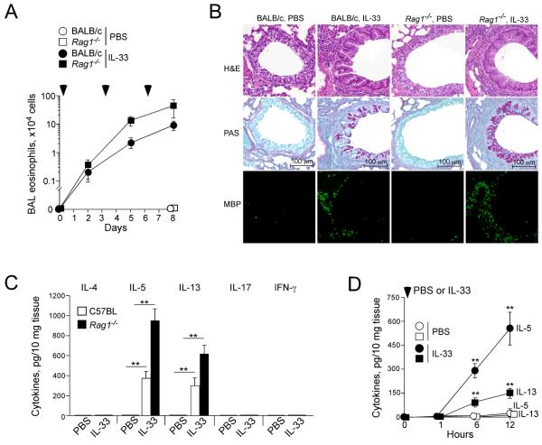
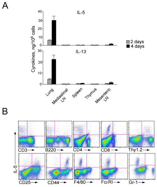
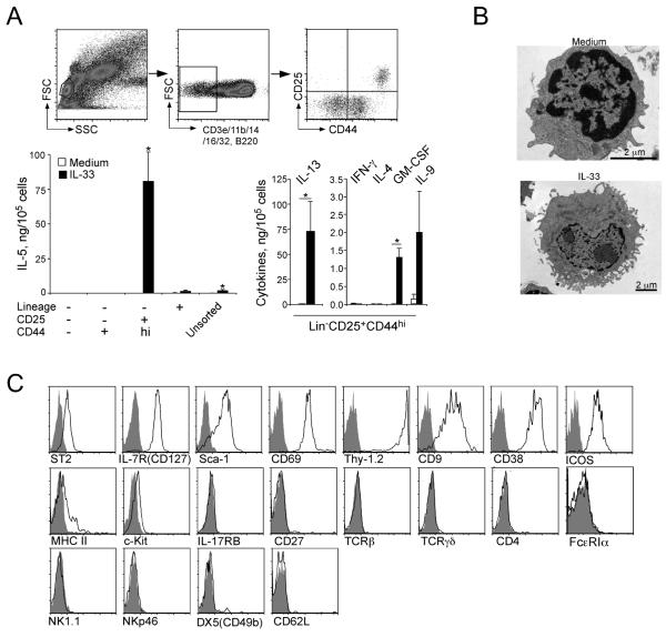
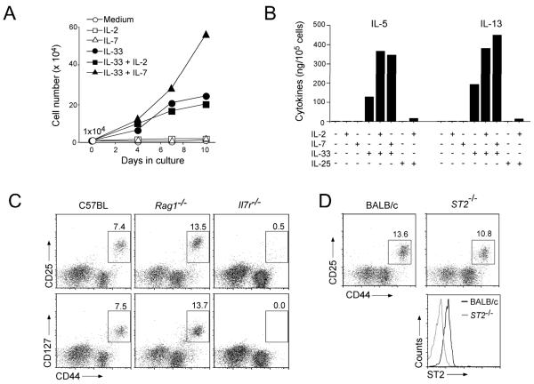
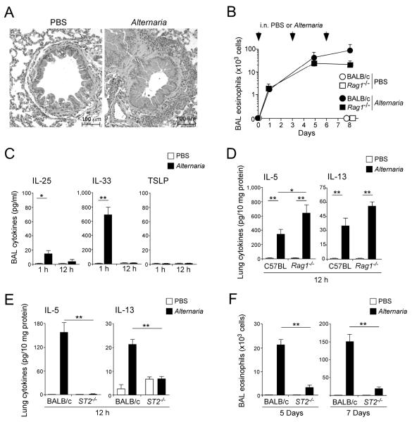
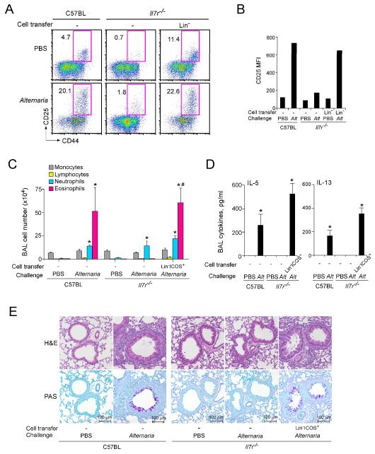
Innate immunity provides the first line of response to invading pathogens and a variety of environmental insults. Recent studies identified novel subsets of innate lymphoid cells that are capable of mediating immune responses in mucosal organs. In this paper, we describe a subset of lymphoid cells that is involved in innate type 2 immunity in the lungs. Airway exposure of naive BALB/c or C57BL/6J mice to IL-33 results in a rapid (<12 h) production of IL-5 and IL-13 and marked airway eosinophilia independently of adaptive immunity. In the lungs of nonsensitized naive mice, IL-33-responsive cells were identified that have a lymphoid morphology, lack lineage markers, highly express CD25, CD44, Thy1.2, ICOS, Sca-1, and IL-7Rα (i.e., Lin(-)CD25(+)CD44(hi) lymphoid cells), and require IL-7Rα for their development. Airway exposure of naive mice to a clinically relevant ubiquitous fungal allergen, Alternaria alternata, increases bronchoalveolar lavage levels of IL-33, followed by IL-5 and IL-13 production and airway eosinophilia without T or B cells. This innate type 2 response to the allergen is nearly abolished in mice deficient in IL-33R (i.e., ST2), and the Lin(-)CD25(+)CD44(hi) lymphoid cells in the lungs are required and sufficient to mediate the response. Thus, a subset of innate immune cells that responds to IL-33 and vigorously produces Th2-type cytokines is present in mouse lungs. These cells may provide a novel mechanism for type 2 immunity in the airways and induction of allergic airway diseases such as asthma.


Figures

Figure 1
Figure 1
IL-33 induces airway eosinophilia and Th2 cytokine production in naïve mice lacking adaptive immunity. (A) IL-33 (100 ng/dose) or PBS were administered i.n. to naïve BALB/c and Rag1−/− mice three times, as indicated by the arrowheads. BAL fluid was collected 48 h after each administration, and the eosinophil number was counted. Data are means ± SEMs of 3-5 mice per group and representative of 2 individual experiments. (B) Lungs were collected 24 h after the third IL-33 administration as described in A, formalin fixed, paraffin embedded, and stained with H&E (top row), PAS (middle row) or anti-MBP (bottom row). (C) IL-33 (100 ng/dose) or PBS were administered i.n. to naïve C57BL and Rag1−/− mice once. After 12 h, lungs were collected, and the cytokine levels in the lung homogenates were measured. Data shown are means ± SEM of 5-8 mice per group and representative of 3 individual experiments. **, p<0.01 compared to PBS administration in each mouse strain. (D) IL-33 or PBS was administered i.n. to naïve BALB/c mice. Lungs were collected at the indicated time points, and the levels of IL-5 (circles) and IL-13 (squares) in the lung homogenates were measured. Data shown are means ± SEM of 4-8 mice per group and representative of 2 individual experiments. **, p<0.01 compared to PBS administration.
Figure 2
Figure 2
Lung cells cultured with IL-33 produce IL-5 and IL-13 in vitro. (A) Single-cell suspensions were obtained from lungs, spleens, thymi and mediastinal and mesenteric LNs of naive BALB/c mice and cultured for 2 or 4 days with IL-33 (10 ng/ml). The concentrations of IL-5 and IL-13 in the cell-free supernatants were measured. (B) Single cell suspensions of lung cells from naive BALB/c mice were cultured with IL-33 (10 ng/ml) for 48 h and stained for intracellular IL-5 and cell surface markers. Data are representative of 4 different experiments showing similar results.
Figure 3
Figure 3
LinCD25+CD44hi lymphoid cells in the lungs produce a large quantity of IL-5 and IL-13 in response to IL-33 in vitro. (A) Four populations of lung cells, including Lin+ cells, LinCD25CD44 cells, Lin CD25CD44+ cells and LinCD25+CD44hi cells were isolated from naïve BALB/c mice by FACS sorting; the upper panels show the gating strategy. Sorted and unsorted lung cells were cultured with medium alone or with 10 ng/ml IL-33 for 96 h, and the levels of cytokines in the supernatants were measured by ELISA. Data shown are means ± SEMs of 4-9 independent experiments. *, p<0.05 compared to cells cultured with medium alone. (B) FACS-sorted LinCD25+CD44hi lung cells were cultured with medium alone or 10 ng/ml IL-33 for 96 h and examined under electron microscopy. Original magnifications; 25,000x (medium alone, top) and 12,000x (IL-33, bottom). (C) Expression of various cell surface markers on the LinCD25+CD44hi lung cell population was examined by flow cytometry. Solid line depicts staining with antibody to the indicated marker; filled grey areas are isotype control staining. Data are representative of three experiments showing similar results.
Figure 4
Figure 4
The roles of IL-2 and IL-7 in proliferation of and cytokine production by Lin CD25+CD44hi lung cells. (A) Isolated LinCD25+CD44hi cells (1×10⁴ cells/well) were cultured with medium alone or with IL-2 (50 ng/ml), IL-7 (10 ng/ml), or IL-33 (10 ng/ml) or their combinations for up to 10 days. Expansion of LinCD25+CD44hi cells was determined by counting the cell number at each time point. Data from one of three experiments showing similar results are presented. (B) Isolated LinCD25+CD44hi cells were cultured with medium alone or with IL-2 (50 ng/ml), IL-7, IL-25, IL-33 (10 ng/ml each) or their combinations for 96 h. The levels of IL-5 and IL-13 in the supernatants were determined by ELISA. Data are representative of three experiments showing similar findings. (C) Single-cells suspensions of lung cells from naive C57BL, Rag1−/− and Il7r−/− mice were stained for lineage markers, CD44, and CD25 or CD127 (IL-7Rα), and Lin lung cells were analyzed as described in Figure 3A. (D) Single-cell suspensions of lung cells from BALB/c and ST2−/− mice were stained with lineage markers, CD44, CD25 and ST2. Top panels: Lin lymphocytic cells were analyzed as described in Figure 3A. Bottom panel: expression of ST2 on LinCD25+CD44hi cells is depicted in BALB/c (black line) and ST2−/− (red line) mice.
Figure 5
Figure 5
Airway exposure of mice to Alternaria extract induces rapid Th2-type cytokine responses through IL-33-dependent innate immune mechanism(s). (A) Naïve BALB/c mice were exposed i.n. three times to Alternaria extract, as described in Materials and Methods. Lungs were collected 24 h after the third exposure, formalin fixed, paraffin embedded and stained with H&E. (B) Naïve BALB/c or Rag1−/− mice were exposed i.n. three times to Alternaria extract or PBS on days 0, 3, and 6. BALs were collected 24 h after the first exposure and 48 h after the second and third exposure, and the eosinophil numbers were determined. Data shown are means ± SEMs of 3-5 mice per group and representative of 3 individual experiments. (C) Naïve BALB/c mice were exposed i.n. once to Alternaria extract or PBS. BAL fluid was collected 1 or 12 h later, and the levels of IL-25, IL-33, and TSLP in the supernatants were determined. Data shown are means ± SEMs of 3-6 mice per group and representative of 3 individual experiments. *, p<0.05; **, p<0.01. (D) Naïve C57BL or Rag1−/− mice were exposed i.n. once to Alternaria extract or PBS. Lungs were collected 12 h later and the levels of IL-5 and IL-13 in lung homogenates were examined. Data shown are means ± SEMs of 4-8 mice per group and representative of 2 individual experiments. *, p<0.05; **, p<0.01. (E) Naïve BALB/c or ST2−/− mice were exposed i.n. once to Alternaria extract or PBS. Lungs were collected 12 h later, and the levels of cytokines in lung homogenates were determined. Data are shown as means ± SEMs of 8-16 mice per group, a pool of 2 individual experiments. **, p<0.01. (F) Naïve BALB/c or ST2−/− mice were exposed i.n. three times to Alternaria extract or PBS as described in B. BAL fluid was collected 48 h after the second exposure (i.e. Day 5) or 24 h after the third exposure (i.e. Day 7), and the numbers of eosinophils were determined. Data are shown as means ± SEMs of 8-16 mice per group from a pool of 2 individual experiments. **, p<0.01.
Figure 6
Figure 6
Lung LinCD25+CD44hi cells are activated when mice are exposed i.n. to IL-33 or Alternaria extract in vivo. (A) Naïve BALB/c mice were exposed i.n. once to PBS, IL-33 or Alternaria extract, and lungs were collected 12 h later. The cell surface expression of CD25 and CD44 on lung Lin cells was analyzed, as described in Figure 2A. One of two experiments with similar results is shown. (B) Left panel: Representative histograms of CD25 intensity in LinCD25+CD44hi cells from mice exposed to PBS (grey filled histogram), IL-33 (green line) or Alternaria (blue line) are shown. Right panel: Mean fluorescence intensity (MFI) of CD25 staining in LinCD25+CD44hi cells was determined from flow cytometry, and data from 3 mice per group are summarized (means ± SEMs). *, p<0.05 compared to PBS administration. (C) Naïve BALB/c or ST2−/− mice were exposed i.n. once to PBS or Alternaria extract, and lungs were collected 12 h later. The cell surface expression of CD25 and CD44 on lung Lin cells was analyzed, as described in Figure 2C. One of two experiments showing similar results is shown. (D) Left panel: Representative histograms of CD25 intensity in LinCD25+CD44hi cells from mice exposed to PBS (BALB/c, grey filled histogram; ST2−/−, dashed black line) or Alternaria (BALB/c, blue line; ST2−/−, green line) are shown. Right panel: MFI of CD25 staining in Lin CD25+CD44hi cells was determined by flow cytometry, and data from 3 mice per group are summarized (means ± SEMs). *, p<0.05 compared to PBS administration.
Figure 7
Figure 7
Adoptive transfer of LinCD25+CD44hi cells from wild type mice reconstitutes the Alternaria-induced immune responses in Il7r−/− mice. (A) Lin cells were isolated from the lungs of naïve C57BL mice as described in Materials and Methods, and they (1×10⁶ cells/mouse) were adoptively transferred intravenously to naive Il7r−/− mice. Control naïve C57BL and Il7r−/− mice received PBS. Twenty-four h later, mice were exposed i.n. once to PBS or Alternaria extract. Lungs were collected 24 h later, and the expression of CD25 and CD44 on lung Lin cells was analyzed, as described in Figure 2C. (B) MFI of CD25 staining in LinCD25+CD44hi cells was determined by flow cytometry. (C) LinICOS+ cells were isolated from the lungs of naïve C57BL mice as described in Materials and Methods, and they (1×10⁵ cells/mouse) were adoptively transferred intravenously to naive Il7r−/− mice. Control naïve C57BL and Il7r−/− mice received PBS. Beginning 24 h later, mice were exposed i.n. three times to PBS or Alternaria extract on days 0, 3, and 6. BAL fluid was collected 24 h after the last exposure, and the cell number and differentials were determined. Data shown are means ± SEMs from 2-4 mice per group. *, p<0.05 compared to the same strain of mice treated with PBS. #, p<0.05 compared to Il7r−/− mice without LinICOS+ cell transfer but exposed to Alternaria extract. (D) The levels of IL-5 and IL-13 in the BAL fluid supernatants of the mice as described in C were determined. *, p<0.05 compared to the same strain of mice treated with PBS. (E) Representative histology (upper panels: H&E staining, lower panels: PAS staining) of the mice as described in C is presented.

References

    1. Robinson DS, Hamid Q, Ying S, Tsicopoulos A, Barkans J, Bentley AM, Corrigan C, Durham SR, Kay AB. Predominant TH2-like bronchoalveolar T-lymphocyte population in atopic asthma. N. Engl. J. Med. 1992;326:298–304. - PubMed
    1. Locksley RM. Asthma and allergic inflammation. Cell. 2010;140:777–783. - PMC - PubMed
    1. Schleimer RP, Kato A, Kern R, Kuperman D, Avila PC. Epithelium: at the interface of innate and adaptive immune responses. J. Allergy Clin. Immunol. 2007;120:1279–1284. - PMC - PubMed
    1. Paul WE, Zhu J. How are T(H)2-type immune responses initiated and amplified? Nat Rev Immunol. 2010;10:225–235. - PMC - PubMed
    1. Wang YH, Liu YJ. Thymic stromal lymphopoietin, OX40-ligand, and interleukin-25 in allergic responses. Clin. Exp. Allergy. 2009;39:798–806. - PMC - PubMed

Publication types