

Atypical extraventricular neurocytoma
- PMID: 22200023
- PMCID: PMC3243844
- DOI: 10.3340/jkns.2011.50.4.381
Atypical extraventricular neurocytoma
Abstract
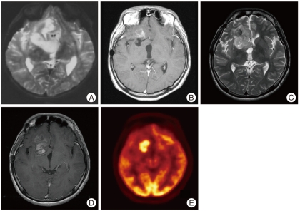
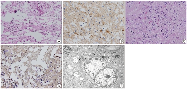
The authors report a case of atypical extraventricular neurocytoma (EVN) transformed from EVN which had been initially diagnosed as an oligodendroglioma 15 years ago. An 8-year-old boy underwent a surgical resection for a right frontal mass which was initially diagnosed as oligodendroglioma. When the tumor recurred 15 years later, a secondary operation was performed, followed by salvage gamma knife treatment. The recurrent tumor was diagnosed as an atypical EVN. The initial specimen was reviewed and immunohistochemistry revealed a strong positivity for synaptophysin. The diagnosis of the initial tumor was revised as an EVN. The patient maintained a stable disease state for 15 years after the first operation, and was followed up for one year without any complications or disease progression after the second operation. We diagnosed an atypical extraventricular neurocytoma transformed from EVN which had been initially diagnosed as an oligodendroglioma 15 years earlier. We emphasize that EVN should be included in the differential diagnosis of oligodendroglioma.
Keywords: Atypical extraventricular neurocytoma; Differential diagnosis; Oligodendroglioma; Recurrence.
Figures
Similar articles
-
Intraventricular central neurocytoma molecularly defined as extraventricular neurocytoma: a case representing the discrepancy between clinicopathological and molecular classifications.Brain Tumor Pathol. 2023 Oct;40(4):230-234. doi: 10.1007/s10014-023-00469-2. Epub 2023 Sep 11. Brain Tumor Pathol. 2023. PMID: 37695397 Free PMC article.
-
Atypical extraventricular neurocytoma.Pathol Int. 2006 Jan;56(1):25-9. doi: 10.1111/j.1440-1827.2006.01914.x. Pathol Int. 2006. PMID: 16398676
-
Atypical extraventricular neurocytoma in a 3-year-old girl: case report with radiological-pathological correlation.Childs Nerv Syst. 2015 Jul;31(7):1189-93. doi: 10.1007/s00381-015-2706-6. Epub 2015 Apr 19. Childs Nerv Syst. 2015. PMID: 25894757
-
[Atypical cerebellar neurocytoma resembling a hemangioblastoma. A case report].Neurocirugia (Astur). 2014 Jul-Aug;25(4):189-93. doi: 10.1016/j.neucir.2014.04.001. Epub 2014 May 16. Neurocirugia (Astur). 2014. PMID: 24837842 Review. Spanish.
-
Imaging of extraventricular neurocytoma: a systematic literature review.Radiol Med. 2020 Oct;125(10):961-970. doi: 10.1007/s11547-020-01198-8. Epub 2020 Apr 25. Radiol Med. 2020. PMID: 32335813
Cited by
-
Congenital Extra-Ventricular (Ganglio)Neurocytoma of the Brain Stem: A Case Report.Front Pediatr. 2018 May 11;6:108. doi: 10.3389/fped.2018.00108. eCollection 2018. Front Pediatr. 2018. PMID: 29868519 Free PMC article.
-
A clinicopathologic study of extraventricular neurocytoma.J Neurooncol. 2017 Mar;132(1):75-82. doi: 10.1007/s11060-016-2336-1. Epub 2016 Nov 18. J Neurooncol. 2017. PMID: 27864704
-
Extraventricular Neurocytoma: Clinical Investigation of Heterogenous Prognosis.Brain Tumor Res Treat. 2022 Jan;10(1):22-28. doi: 10.14791/btrt.2022.10.e30. Brain Tumor Res Treat. 2022. PMID: 35118844 Free PMC article.
-
Extraventricular neurocytoma in the left temporal lobe: A case report and review of the literature.Oncol Lett. 2016 Jun;11(6):3579-3582. doi: 10.3892/ol.2016.4487. Epub 2016 Apr 21. Oncol Lett. 2016. PMID: 27313678 Free PMC article.
-
Extraventricular neurocytoma of the sellar region: case report and literature review.Springerplus. 2016 Jul 7;5(1):987. doi: 10.1186/s40064-016-2650-2. eCollection 2016. Springerplus. 2016. PMID: 27398267 Free PMC article.
References
-
- Brandes AA, Amistà P, Gardiman M, Volpin L, Danieli D, Guglielmi B, et al. Chemotherapy in patients with recurrent and progressive central neurocytoma. Cancer. 2000;88:169–174. - PubMed
-
- Brat DJ, Scheithauer BW, Eberhart CG, Burger PC. Extraventricular neurocytomas : pathologic features and clinical outcome. Am J Surg Pathol. 2001;25:1252–1260. - PubMed
-
- Choi YS, Song YJ, Huh KY, Kim KU. Malignant variant of the central neurocytoma. J Korean Neurosurg Soc. 2004;35:313–316.
-
- Fujisawa H, Marukawa K, Hasegawa M, Tohma Y, Hayashi Y, Uchiyama N, et al. Genetic differences between neurocytoma and dysembryoplastic neuroepithelial tumor and oligodendroglial tumors. J Neurosurg. 2002;97:1350–1355. - PubMed
Publication types
LinkOut - more resources
Full Text Sources
