

Case Reports
doi: 10.3340/jkns.2011.50.4.385.
Epub 2011 Oct 31.
Malignant ascites after subduroperitoneal shunt in a patient with leptomeningeal metastasis
Affiliations
- PMID: 22200024
- PMCID: PMC3243845
- DOI: 10.3340/jkns.2011.50.4.385
Item in Clipboard
Case Reports
Malignant ascites after subduroperitoneal shunt in a patient with leptomeningeal metastasis
J Korean Neurosurg Soc.
2011 Oct.
Abstract
Leptomeningeal metastasis is a devastating complication of advanced stage cancer. It is frequently accompanied by hydrocephalus and intracranial hypertension that must be treated by ventriculoperitoneal shunts. However, there are actual risks of peritoneal seeding or accumulation of malignant ascites after the cerebrospinal fluid diversion procedure, though it has not been reported. Here, we present the case of a patient with non-small cell lung cancer with leptomeningeal metastasis in whom malignant ascites developed after a subduroperitoneal shunt.
Keywords: Leptomeningeal metastasis; Malignant ascites; Ventriculoperitoneal shunt.
Figures

Axial T1-weighted contarst-enhanced MRI shows subdural fluid collection around the left cerebral hemisphere resulting in midline shift and asymmetrical ventricles. Also, tissue defect from previous surgical resection of the metastatic lesion is shown in left parietal area. MRI : magnetic resonance image.
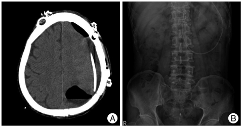
A : Brain CT scan after subduroperitoneal shunt shows a subdural catheter around the left cerebral hemisphere and demonstrates communication between subdural space and previous tumor resection cavity by pneumocephalus in both space. B : Plain X-ray image of abdomen shows a distal shunt catheter within peritoneal cavity. CT : computed tomography.
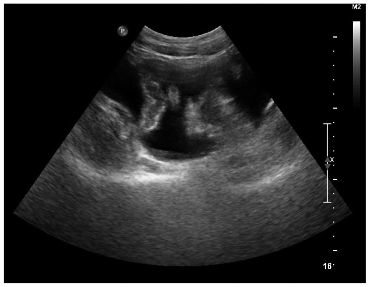
Ultrasonography reveals a large amount of ascites in the abdominal and pelvic cavity.
Similar articles
-
Palliative lumboperitoneal shunt for leptomeningeal metastasis-related hydrocephalus: A case series.Palliat Med. 2017 Jan;31(1):93-96. doi: 10.1177/0269216316649128. Epub 2016 Jul 10. Palliat Med. 2017. PMID: 27188875
-
Childhood optic pathway tumors associated with ascites following ventriculoperitoneal shunt placement.Pediatr Neurosurg. 1994;21(4):254-8; discussion 259. doi: 10.1159/000120846. Pediatr Neurosurg. 1994. PMID: 7865412
-
Lumboperitoneal shunt for the treatment of leptomeningeal metastasis.Med Hypotheses. 2015 May;84(5):506-8. doi: 10.1016/j.mehy.2015.02.009. Epub 2015 Feb 25. Med Hypotheses. 2015. PMID: 25754849
-
Diffuse midline glioma metastasis to the peritoneal cavity via ventriculo-peritoneal shunt: Case report and review of literature.J Clin Neurosci. 2019 Sep;67:288-293. doi: 10.1016/j.jocn.2019.06.043. Epub 2019 Jun 29. J Clin Neurosci. 2019. PMID: 31266714 Review.
-
[Ascites of the cerebrospinal fluid as a complication of a ventriculoperitoneal shunt: report of a case and a review of the literature].An Esp Pediatr. 1988 Jun;28(6):565-8. An Esp Pediatr. 1988. PMID: 3057973 Review. Spanish.
Cited by
-
Leptomeningeal disease in melanoma patients: An update to treatment, challenges, and future directions.Pigment Cell Melanoma Res. 2020 Jul;33(4):527-541. doi: 10.1111/pcmr.12861. Epub 2020 Jan 19. Pigment Cell Melanoma Res. 2020. PMID: 31916400 Free PMC article. Review.
-
Improved Survival and Symptom Relief Following Palliative Cerebrospinal Fluid Diversion for Leptomeningeal Disease from Brain Cancers: A Case Series and Systematic Review.Cancers (Basel). 2025 Jan 17;17(2):292. doi: 10.3390/cancers17020292. Cancers (Basel). 2025. PMID: 39858073 Free PMC article.
-
Ventriculoperitoneal shunting with an on-off valve for patients with leptomeningeal metastases and intracranial hypertension.Neurooncol Pract. 2023 Sep 2;11(1):56-63. doi: 10.1093/nop/npad056. eCollection 2024 Feb. Neurooncol Pract. 2023. PMID: 38222058 Free PMC article.
References
-
- Chamberlain MC. Leptomeningeal metastases : a review of evaluation and treatment. J Neurooncol. 1998;37:271–284. - PubMed
-
- Coelho Neto M, Ramina R, de Meneses MS, Arruda WO, Milano JB. Peritoneal dissemination from central neurocytoma : case report. Arq Neuropsiquiatr. 2003;61:1030–1034. - PubMed
-
- Eralp Y, Saip P, Aydin Z, Berkman S, Topuz E. Leptomeningeal dissemination of ovarian carcinoma through a ventriculoperitoneal shunt. Gynecol Oncol. 2008;108:248–250. - PubMed
-
- Glantz MJ, Jaeckle KA, Chamberlain MC, Phuphanich S, Recht L, Swinnen LJ, et al. A randomized controlled trial comparing intrathecal sustained-release cytarabine (DepoCyt) to intrathecal methotrexate in patients with neoplastic meningitis from solid tumors. Clin Cancer Res. 1999;5:3394–3402. - PubMed
-
- Herrlinger U, Förschler H, Küker W, Meyermann R, Bamberg M, Dichgans J, et al. Leptomeningeal metastasis : survival and prognostic factors in 155 patients. J Neurol Sci. 2004;223:167–178. - PubMed
Publication types
LinkOut - more resources
Full Text Sources
