. 2011 Dec 26:8:183.
doi: 10.1186/1742-2094-8-183.

Interleukin-1 beta and neurotrophin-3 synergistically promote neurite growth in vitro

Affiliations

Interleukin-1 beta and neurotrophin-3 synergistically promote neurite growth in vitro

Francesco Boato et al. J Neuroinflammation. .

Abstract

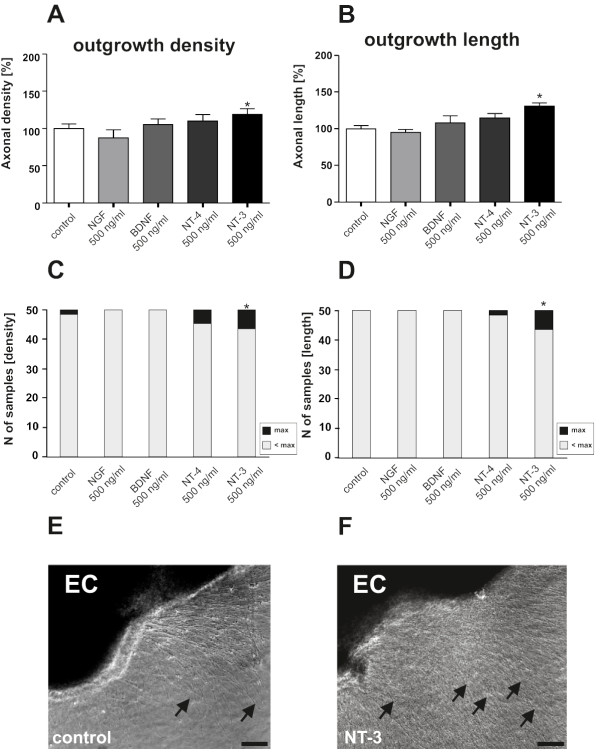
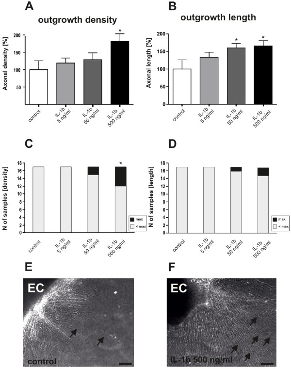
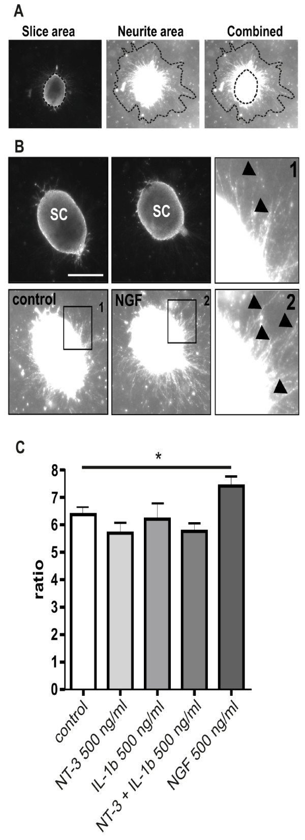
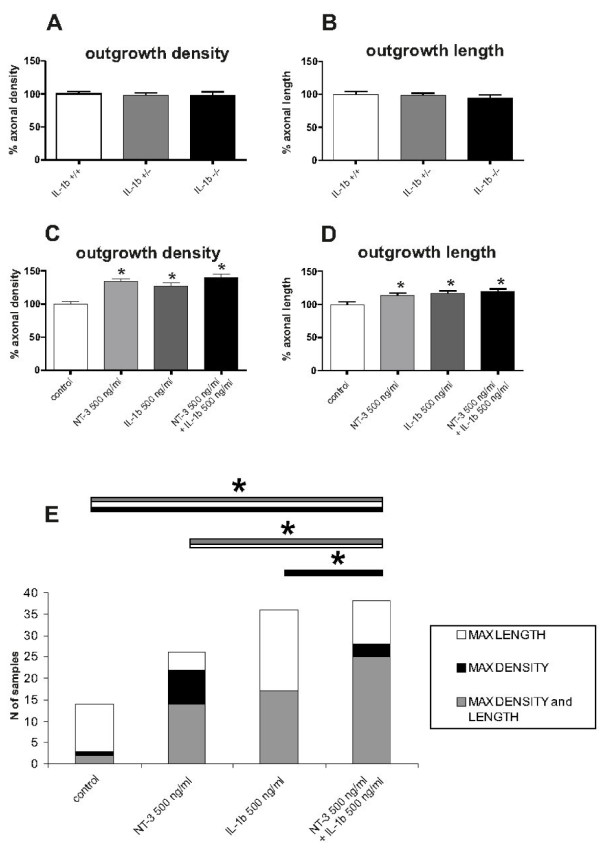
Pro-inflammatory cytokines such as interleukin-1 beta (IL-1β) are considered to exert detrimental effects during brain trauma and in neurodegenerative disorders. Consistently, it has been demonstrated that IL-1β suppresses neurotrophin-mediated neuronal cell survival rendering neurons vulnerable to degeneration. Since neurotrophins are also well known to strongly influence axonal plasticity, we investigated here whether IL-1β has a similar negative impact on neurite growth. We analyzed neurite density and length of organotypic brain and spinal cord slice cultures under the influence of the neurotrophins NGF, BDNF, NT-3 and NT-4. In brain slices, only NT-3 significantly promoted neurite density and length. Surprisingly, a similar increase of neurite growth was induced by IL-1β. Additionally, both factors increased the number of brain slices displaying maximal neurite growth. Furthermore, the co-administration of IL-1β and NT-3 significantly increased the number of brain slices displaying maximal neurite growth compared to single treatments. These data indicate that these two factors synergistically stimulate two distinct aspects of neurite outgrowth, namely neurite density and neurite length from acute organotypic brain slices.


Figures

Figure 1
Figure 1
Recombinant NT-3 stimulates neurite density and length of organotypic brain slices. The neurotrophins NGF, BDNF, NT-3 and NT-4 (500 ng/ml) were added to the culture medium immediately after preparation of the organotypic brain slices. NT-3, but not the other neurotrophins significantly increases neurite density (A), neurite length (B), the amount of slices reaching the maximum outgrowth (C) and the amount of slices reaching the maximum length (D). E + F: representative photomicrograph showing the increase in outgrowth of NT-3 treated EC slices compared to control. n = 50 slices. A + B: *: Statistically significant difference to control; p < 0.05 (Mann Whitney U test). C + D: *: Statistically significant difference; p < 0.05 (Chi-square analysis). EC = enthorinal cortex. Arrows indicate outgrowing neuritis. Scale bar: 100 μm.
Figure 2
Figure 2
IL-1β stimulates neurite density and length in organotypic brain slices. A dose-response curve revealed that high doses of IL-1β (500 ng/ml) added to the culture medium, stimulate neurite density (A) of organotypic brain slices and the amount of slices reaching the maximum outgrowth (C). A lower dose (50 ng/ml) is still able to stimulate the average length of neuritis (B) but neither 500 ng/ml nor 50 ng/ml of IL-1β were able to significantly increase the amount of slices presenting maximum length (D). E + F: representative photomicrograph showing the increase in outgrowth of IL-1β treated EC compared to control. n = 17 slices. A + B: *: Statistically significant difference to control; p < 0.05 (Mann Whitney U test). C + D: *: Statistically significant difference; p < 0.05 (Chi-square analysis) EC = enthorinal cortex. Arrows indicate neuritis. Scale bar: 100 μm.
Figure 3
Figure 3
NT-3 and IL-1beta do not increase neurite outgrowth of transverse spinal cord slices. A: Transverse spinal cord slices were prepared from E13 spinal cords and the ratio between neurite area and slice area were compared. B: representative photomicrograph showing the increase in outgrowth of NGF treated EC compared to control. NGF (500 ng/ml) serves as positive control. C: NT-3 and IL-1β (500 ng/ml) were added to the culture medium of organotypic transverse spinal cord slices. Only NGF significantly increases neurite density, while NT-3, IL-1β or a combination of these factors does not influence neurite outgrowth. n = 9-11 slices. *: Statistically significant difference to control; p < 0.05 (Mann Whitney U test). Arrow heads indicate outgrowing neuritis. Scale bar: 50 μm.
Figure 4
Figure 4
Neurite outgrowth is independent on endogenous IL-1beta and is synergistically stimulated by combined application of NT-3 and IL-1beta. A + B: Neurite outgrowth (neurite density A and neurite length B) was not influenced in the absence of endogenous IL-1β in IL-1β-deficient mice. Heterozygous IL-1β-deficient and wildtype mice served as controls. n: 50 slices. C + D: The combined administration of NT-3 and IL-1β shows only a slight increase in neurite density and length, if compared to single treatments. n = 84 slices. *: Statistically significant difference to control; p < 0.05 (Mann Whitney U test). Error bars represent SEM. E: Chi-square analysis reveals a significant difference in the frequency of brain slices with maximal outgrowth between single treatments with NT3 or IL-1β and the combined administration of both factors. Particularly double treatment presents a significant increase in the number of slices reaching maximum density if compared to control and single treatment with IL-1β, and in length if compared to single treatment with NT3. Additionally, combination of the two factors is also characterized by a significantly higher number of slices which hit the maximum in both parameters, if compared to control condition or single treatment with NT3 (to increase the readability of the graph only the significances relative to the double treatment have been included). *: Statistically significant difference; p < 0.05 (Chi-square).

Similar articles

Cited by

References

    1. Peschke T, Bender A, Nain M, Gemsa D. Role of macrophage cytokines in influenza A virus infections. Immunobiology. 1993;189(3-4):340–355. doi: 10.1016/S0171-2985(11)80365-7. - DOI - PubMed
    1. Hildebrand F, Pape HC, Krettek C. The importance of cytokines in the posttraumatic inflammatory reaction. Unfallchirurg. 2005;108(10):793–794, 796-803. doi: 10.1007/s00113-005-1005-1. - DOI - PubMed
    1. Bauer J, Berkenbosch F, Van Dam AM, Dijkstra CD. Demonstration of interleukin-1 beta in Lewis rat brain during experimental allergic encephalomyelitis by immunocytochemistry at the light and ultrastructural level. Journal of neuroimmunology. 1993;48(1):13–21. doi: 10.1016/0165-5728(93)90053-2. - DOI - PubMed
    1. Boutin H, LeFeuvre RA, Horai R, Asano M, Iwakura Y, Rothwell NJ. Role of IL-1alpha and IL-1beta in ischemic brain damage. J Neurosci. 2001;21(15):5528–5534. - PMC - PubMed
    1. Loddick SA, Rothwell NJ. Neuroprotective effects of human recombinant interleukin-1 receptor antagonist in focal cerebral ischaemia in the rat. J Cereb Blood Flow Metab. 1996;16(5):932–940. - PubMed

Publication types