Review
. 2012 Nov;238(1):1-11.
doi: 10.1016/j.expneurol.2011.12.013. Epub 2011 Dec 19.

Protein aggregates in Huntington's disease

Affiliations
Review

Protein aggregates in Huntington's disease

Montserrat Arrasate et al. Exp Neurol. 2012 Nov.

Abstract

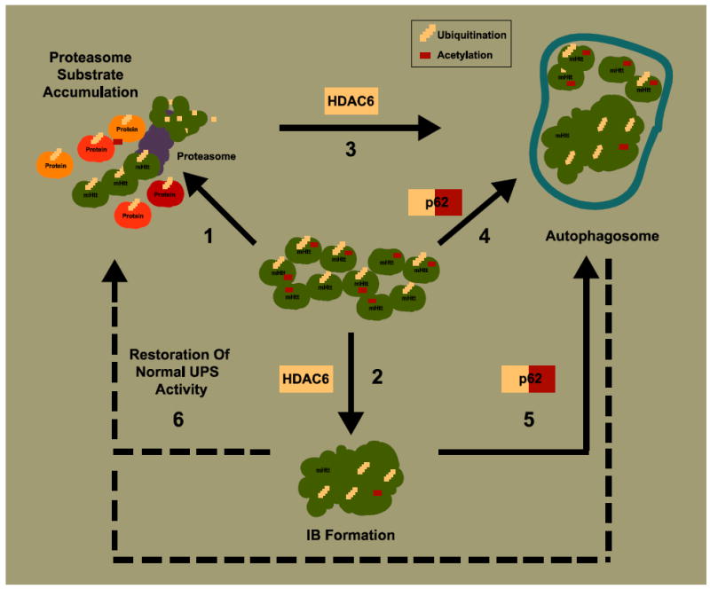
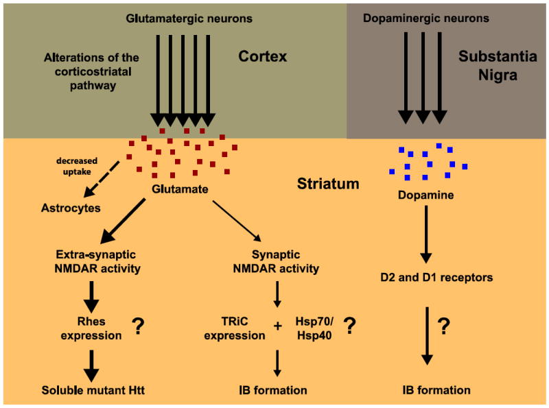
Huntington's disease (HD) is an incurable neurodegenerative disease characterized by abnormal motor movements, personality changes, and early death. HD is caused by a mutation in the IT-15 gene that expands abnormally the number of CAG nucleotide repeats. As a result, the translated protein huntingtin contains disease-causing expansions of glutamines (polyQ) that make it prone to misfold and aggregate. While the gene and mutations that cause HD are known, the mechanisms underlying HD pathogenesis are not. Here we will review the state of knowledge of HD, focusing especially on a hallmark pathological feature-intracellular aggregates of mutant Htt called inclusion bodies (IBs). We will describe the role of IBs in the disease. We speculate that IB formation could be just one component of a broader coping response triggered by misfolded Htt whose efficacy may depend on the extent to which it clears toxic forms of mutant Htt. We will describe how IB formation might be regulated and which factors could determine different coping responses in different subsets of neurons. A differential regulation of IB formation as a function of the cellular context could, eventually, explain part of the neuronal vulnerability observed in HD.


Figures

Figure 1
Figure 1. Model for cellular regulation of coping responses to the presence of misfolded mutant Htt
(1) Expression of mutant Htt causes accumulation of substrates requiring proteasome degradation. This might be due to an increase in the amounts of other misfolded proteins that compete for proteasomal degradation or to impairment of proteasome function or both. “Sensor” proteins that recognize ubiquitinated and acetylated forms of mutant Htt, such as HDAC6 and p62, might facilitate IB formation and autophagy activation. (2) HDAC6 binds ubiquitin and favors accumulation of ubiquitinated protein into IBs. (3) HDAC6 also induces autophagy as a response to polyQ-dependent proteasome substrate accumulation. (4) p62 interacts with acetylated mutant Htt potentially targeting the protein to degradation by autophagy. (5) p62 also recognizes ubiquitin and might recruit autophagosomal components to degrade non-aggregated and aggregated forms of ubiquitinated mutant Htt. (6) IB formation and autophagy induction would reduce the load of misfolded mutant Htt protein and potentially restore normal ubiquitin proteasome system (UPS) activity.
Figure 2
Figure 2. Potential non-cell-autonomous mechanisms for regulating IB formation in the striatum
Glutamatergic and dopaminergic neuronal projections are the main afferents to the striatum from the cortex and the substantia nigra, respectively. (Left) Extra-synaptic and synaptic activity mediated by glutamate could modulate IB formation. An excess of glutamatergic stimulation might occur in the striatum of HD brains through different mechanisms: increased release from cortical afferents, decreased uptake by striatal astrocytes, and changes in NMDA receptor (NMDAR) number/subunit composition and localization. Excessive extra-synaptic NMDAR-mediated activity might drive gene expression that stabilizes soluble toxic forms of mutant Htt (thicker arrows), leading to neuronal death. Pharmacological blockage of extra-synaptic NMDAR activity would eventually restore an appropriated balance between extra-synaptic and synaptic activity, favoring gene expression that would increase IB formation and neuroprotection. (Right) Dopamine might also modulate IB formation in the striatum through D2 and/or D1 receptors, via unknown mechanisms.

Similar articles

Cited by

References

    1. Aiken CT, Steffan JS, Guerrero CM, Khashwji H, Lukacsovich T, Simmons D, Purcell JM, Menhaji K, Zhu YZ, Green K, Laferla F, Huang L, Thompson LM, Marsh JL. Phosphorylation of threonine 3: implications for Huntingtin aggregation and neurotoxicity. J Biol Chem. 2009;284:29427–29436. - PMC - PubMed
    1. Arrasate M, Finkbeiner S. Automated microscope system for determining factors that predict neuronal fate. Proc Natl Acad Sci U S A. 2005;102:3840–3845. - PMC - PubMed
    1. Arrasate M, Mitra S, Schweitzer ES, Segal MR, Finkbeiner S. Inclusion body formation reduces levels of mutant huntingtin and the risk of neuronal death. Nature. 2004;431:805–810. - PubMed
    1. Atwal RS, Desmond CR, Caron N, Maiuri T, Xia J, Sipione S, Truant R. Kinase inhibitors modulate huntingtin cell localization and toxicity. Nat Chem Biol. 2011;7:453–460. - PubMed
    1. Becher MW, Kotzuk JA, Sharp AH, Davies SW, Bates GP, Price DL, Ross CA. Intranuclear neuronal inclusions in Huntington’s disease and dentatorubral and pallidoluysian atrophy: correlation between the density of inclusions and IT15 CAG triplet repeat length. Neurobiol Dis. 1998;4:387–397. - PubMed

Publication types