

Characterization of oxaliplatin-induced chronic painful peripheral neuropathy in the rat and comparison with the neuropathy induced by paclitaxel
- PMID: 22200546
- PMCID: PMC3273641
- DOI: 10.1016/j.neuroscience.2011.12.023
Characterization of oxaliplatin-induced chronic painful peripheral neuropathy in the rat and comparison with the neuropathy induced by paclitaxel
Abstract
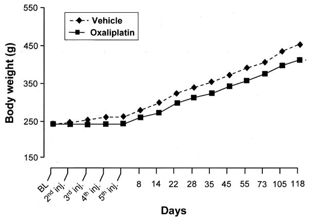
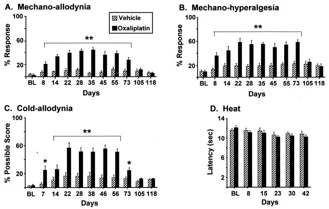
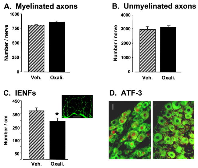
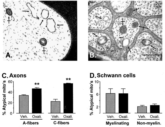
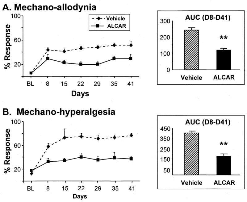
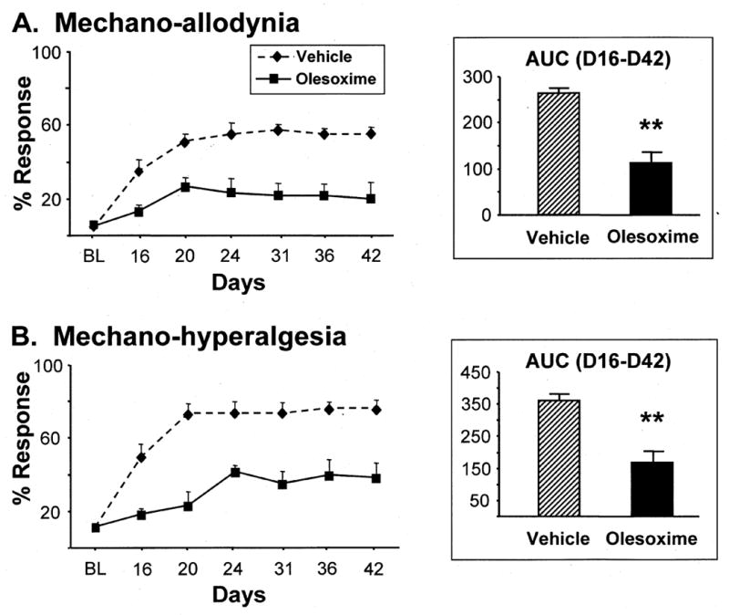
Anti-neoplastic agents in the platinum-complex, taxane, vinca alkaloid, and proteasome-inhibitor classes induce a dose-limiting, chronic, distal, symmetrical, sensory peripheral neuropathy that is often accompanied by neuropathic pain. Clinical descriptions suggest that these conditions are very similar, but clinical data are insufficient to determine the degree of similarity and to determine if they share common pathophysiological mechanisms. Animal models do not have the limitations of clinical studies and so we have characterized a rat model of chronic painful peripheral neuropathy induced by a platinum-complex agent, oxaliplatin, in order to compare it with a previously characterized model of chronic painful peripheral neuropathy induced by a taxane agent, paclitaxel. The oxaliplatin model evokes mechano-allodynia, mechano-hyperalgesia, and cold-allodynia that have a delayed onset, gradually increasing severity, a distinct delay to peak severity, and duration of about 2.5 months. There is no effect on heat sensitivity. Electron microscopy (EM) analyses found no evidence for axonal degeneration in peripheral nerve, and there is no upregulation of activating transcription factor-3 in the lumbar dorsal root ganglia. There is a statistically significant loss of intraepidermal nerve fibers in the plantar hind paw skin. Oxaliplatin treatment causes a significant increase in the incidence of swollen and vacuolated mitochondria in peripheral nerve axons, but not in their Schwann cells. Nerve conduction studies found significant slowing of sensory axons, but no change in motor axons. Single fiber recordings found an abnormal incidence of A- and C-fibers with irregular, low-frequency spontaneous discharge. Prophylactic dosing with two drugs that are known to protect mitochondria, acetyl-l-carnitine and olesoxime, significantly reduced the development of pain hypersensitivity. Our results are very similar to those obtained previously with paclitaxel, and support the hypothesis that these two agents, and perhaps other chemotherapeutics, produce very similar conditions because they have a mitotoxic effect on primary afferent neurons.
Copyright © 2011 IBRO. Published by Elsevier Ltd. All rights reserved.
Conflict of interest statement
None of the authors has any conflict of interest with respect to the contents of this report.
Figures
Similar articles
-
Effects of mitochondrial poisons on the neuropathic pain produced by the chemotherapeutic agents, paclitaxel and oxaliplatin.Pain. 2012 Mar;153(3):704-709. doi: 10.1016/j.pain.2011.12.011. Epub 2012 Jan 13. Pain. 2012. PMID: 22244441 Free PMC article.
-
Mitochondrial abnormality in sensory, but not motor, axons in paclitaxel-evoked painful peripheral neuropathy in the rat.Neuroscience. 2011 Dec 29;199:461-9. doi: 10.1016/j.neuroscience.2011.10.010. Epub 2011 Oct 19. Neuroscience. 2011. PMID: 22037390 Free PMC article.
-
Functional deficits in peripheral nerve mitochondria in rats with paclitaxel- and oxaliplatin-evoked painful peripheral neuropathy.Exp Neurol. 2011 Dec;232(2):154-61. doi: 10.1016/j.expneurol.2011.08.016. Epub 2011 Aug 30. Exp Neurol. 2011. PMID: 21907196 Free PMC article.
-
Bioenergetic deficits in peripheral nerve sensory axons during chemotherapy-induced neuropathic pain resulting from peroxynitrite-mediated post-translational nitration of mitochondrial superoxide dismutase.Pain. 2013 Nov;154(11):2432-2440. doi: 10.1016/j.pain.2013.07.032. Epub 2013 Jul 25. Pain. 2013. PMID: 23891899 Free PMC article.
-
[Role of Transient Receptor Potential Channels in Paclitaxel- and Oxaliplatin-induced Peripheral Neuropathy].Yakugaku Zasshi. 2016;136(2):287-96. doi: 10.1248/yakushi.15-00214. Yakugaku Zasshi. 2016. PMID: 26831807 Review. Japanese.
Cited by
-
Impaired microvascular reactivity in patients treated with 5-fluorouracil chemotherapy regimens: Potential role of endothelial dysfunction.Int J Cardiol Heart Vasc. 2023 Nov 17;49:101300. doi: 10.1016/j.ijcha.2023.101300. eCollection 2023 Dec. Int J Cardiol Heart Vasc. 2023. PMID: 38173789 Free PMC article.
-
Sustained Accumulation of Microtubule-Binding Chemotherapy Drugs in the Peripheral Nervous System: Correlations with Time Course and Neurotoxic Severity.Cancer Res. 2016 Jun 1;76(11):3332-9. doi: 10.1158/0008-5472.CAN-15-2525. Epub 2016 Apr 13. Cancer Res. 2016. PMID: 27197173 Free PMC article.
-
Protective effect of a mitochondria-targeting peptide against paclitaxel-induced peripheral neuropathy.Chem Biol Drug Des. 2023 Apr;101(4):1012-1018. doi: 10.1111/cbdd.14192. Epub 2022 Dec 23. Chem Biol Drug Des. 2023. PMID: 36513390 Free PMC article.
-
Role of non-macrophage cell-derived HMGB1 in oxaliplatin-induced peripheral neuropathy and its prevention by the thrombin/thrombomodulin system in rodents: negative impact of anticoagulants.J Neuroinflammation. 2019 Oct 30;16(1):199. doi: 10.1186/s12974-019-1581-6. J Neuroinflammation. 2019. PMID: 31666085 Free PMC article.
-
Pathobiology of cancer chemotherapy-induced peripheral neuropathy (CIPN).Front Pharmacol. 2013 Dec 18;4:156. doi: 10.3389/fphar.2013.00156. Front Pharmacol. 2013. PMID: 24385965 Free PMC article. Review.
References
-
- Adelsberger H, Quasthoff S, Grosskruetuz J, Lepier A, Eckel F, Lersch C. The chemotherapeutic oxaliplatin alters voltage-gated Na(+) channel kinetics on rat sensory neurons. Eur J Pharmacol. 2000;406:25–32. - PubMed
-
- Ames BN, Liu J. Delaying the mitochondrial decay of aging with acetylcarnitine. Ann N Y Acad Sci. 2004;1033:108–116. - PubMed
-
- Argyriou AA, Polychronopoulos P, Iconomou G, Chroni E, Kalofonos HP. A review on oxaliplatin-induced peripheral nerve damage. Cancer Treat Rev. 2008;34:368–377. - PubMed
-
- Augusto C, Pietro M, Cinzia M, Sergio C, Sara C, Luca G, Scaioli V. Peripheral neuropathy due to paclitaxel: study of the temporal relationships between the therapeutic schedule and the clinical quantitative score (QST) and comparison with neurophysiological findings. J Neurooncol. 2008;86:89–99. - PubMed
Publication types
MeSH terms
Substances
Grants and funding
LinkOut - more resources
Full Text Sources
Other Literature Sources
