

doi: 10.1016/j.canlet.2011.12.027.
Epub 2011 Dec 23.
Interferon Regulatory Factor 1 (IRF-1) induces p21(WAF1/CIP1) dependent cell cycle arrest and p21(WAF1/CIP1) independent modulation of survivin in cancer cells
Affiliations
- PMID: 22200613
- PMCID: PMC3304016
- DOI: 10.1016/j.canlet.2011.12.027
Item in Clipboard
Interferon Regulatory Factor 1 (IRF-1) induces p21(WAF1/CIP1) dependent cell cycle arrest and p21(WAF1/CIP1) independent modulation of survivin in cancer cells
Cancer Lett.
.
Abstract
We have shown that the ectopic expression of Interferon Regulatory Factor 1 (IRF-1) results in human cancer cell death accompanied by the down-regulation of the Inhibitor of Apoptosis Protein (IAP) survivin and the induction of the cyclin-dependent kinase inhibitor p21(WAF1/CIP1). In this report, we investigated the direct role of p21 in the suppression of survivin. We show that IRF-1 down-regulates cyclin B1, cdc-2, cyclin E, E2F1, Cdk2, Cdk4, and results in p21-mediated G1 cell cycle arrest. Interestingly, while p21 directly mediates G1 cell cycle arrest, IRF-1 or other IRF-1 signaling pathways may directly regulate survivin in human cancer cells.
Copyright © 2011 Elsevier Ireland Ltd. All rights reserved.
Figures
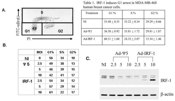
IRF-1 induces a dose-dependent G1 arrest in human breast cancer. (A) MDA-MB-468 human breast cancer cells were either not infected (NI), or infected with the control Ad-Ψ5 or Ad-IRF-1 recombinant adenoviruses at varying multiplicities of infection (MOI) and cell cycle arrest at 24 h was evaluated by FITC-BrdU labeling and propidium iodide staining. An example of the bivariate cell cycle output data is shown. Increases in BrdU are associated with S phase while PI staining indicates either one or two copies of DNA corresponding with G1 or G2 phases. Percentage of cells in each phase can then be calculated. (B) MDA-MB-468 cells were either not infected (NI) or infected with either Ad-Ψ5 or Ad-IRF-1 at the indicated MOIs. Cells were fixed in ETOH, permeabilized, and stained with FITC-αBrdU and propidium iodide (PI). Cell cycle analysis was then performed and percentages of cells in each phase, G1, S, and G2 are shown as assessed by two dimensional analyses as seen in A. (C) MDA-MB-468 cells were either not infected (NI) or infected with either Ad-Ψ5 or Ad-IRF-1 at the indicated MOIs. Infected cells were harvested 24 h after infection, and immunoblotting was conducted as described in Materials and Methods.
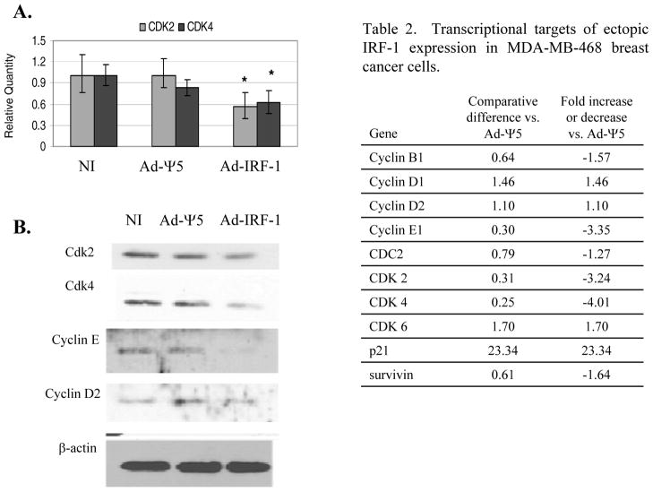
Ectopic expression of IRF-1 suppresses Cdk2, Cdk4, and Cyclin E1 expression. (A) MDA-MB-468 breast cancer cells were either uninfected (NI), or infected with the Ad-Ψ5 control or Ad-IRF-1 adenovirus at MOI 25. Lysates were harvested at 24 hours post infection. Quantitative real-time RT-PCR was performed as described in Materials and Methods, and relative quantities of cdk2 and cdk4 were computed by utilizing the MxPro™ software. These experiments were repeated twice with comparable results. * denotes p<0.05 by ANOVA. (B) MDA-MB-468 breast cancer cells were either uninfected or infected with Ad-Ψ5 control or Ad-IRF-1 at a MOI of 10. Cellular lysates were prepared and Western immunoblotting was conducted as has been described in Materials and Methods. Blots were probed for Cdk2, Cdk4, cyclin E, and β-actin.
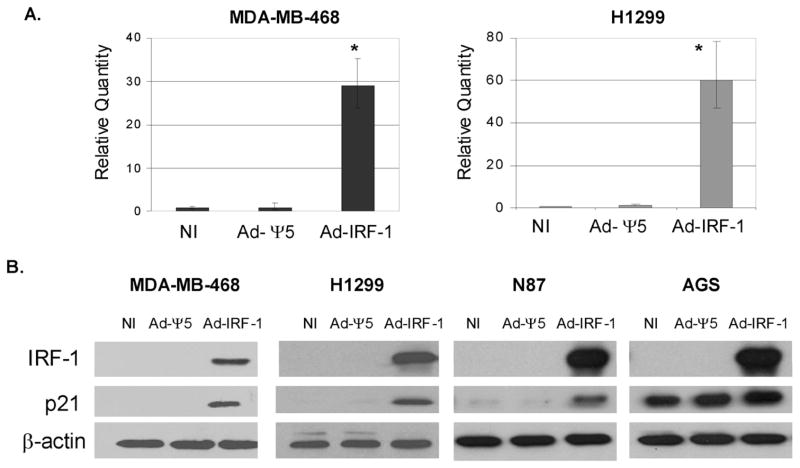
IRF-1 induces the upregulation of p21 mRNA and protein expression in cancer cells. (A) MDA-MB-468 human breast cancer cells and H1299 human non-small-cell lung cancer cells were not infected (NI), or infected with the control Ad-Ψ5 vector or Ad-IRF-1 adenoviruses at MOI 25. 24 h post infection, cell pellets were lysed with TRIzol® and RNA was purified with the Qiagen RNeasy kit. Quantitative real time RT-PCR was performed as described in Materials and Methods. Relative quantities of p21 mRNA were computed by MxPro™ quantitative PCR software (Stratagene, La Jolla, CA). * denotes p<0.005 by ANOVA. (B) MDA-MB-468 and H1299 cancer cell lines were infected as described in A. AGS and N87 gastric cancer cells were infected at MOI 50. 24 h post infection, cells were harvested and immunoblotting was performed as described in Materials and Methods.
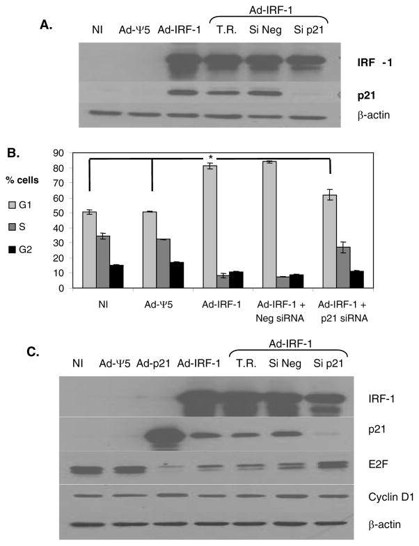
p21 mediates IRF-1 induced G1 arrest in human non-small-cell lung cancer. (A) H1299 lung cancer cells were either untransfected or transfected with a control siNeg, or siRNA to p21. Untransfected cells treated with the transfection reagent (T.R.) siPORT™
Amine are included as an additional control. 24 hours post transfection the cells were either not infected, or infected with Ad-Ψ5 or Ad-IRF-1 recombinant replication defective adenoviruses at a MOI of 25. 24 hours post infection, cells were harvested and immunoblotting was performed as described in Materials and Methods. (B) H1299 cells were treated as in A, and 24 hours post infection, cells were fixed in 80% ETOH, permeabilized, and stained with propidium iodide. Cells were then assayed for cell cycle arrest and these data were analyzed via ModFit LT™ software. ANOVA analysis was utilized to compare the means between no infection NI, Ad-Ψ5, and Ad-IRF-1, while the Student’s t test compared means between Ad-IRF-1 and Ad-IRF-1 + p21 siRNA (* = p<0.05 for both analyses). These experiments were conducted in triplicate. (C) H1299 cells were transfected as in A, and subsequently infected with Ad-Ψ5, Ad-IRF-1, or Ad-p21 at a MOI of 25. Cells were harvested 24 h post infection and immunoblotting was conducted as has been described in Materials and Methods. These experiments were repeated twice with comparable results.
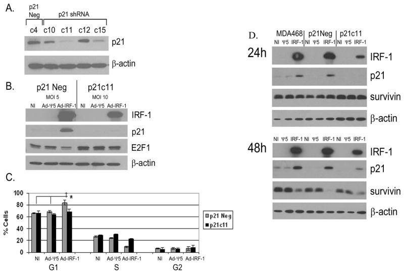
p21 mediates IRF-1 induced G1 arrest but not suppression of survivin in breast cancer cells. (A) MDA-MB-468 cells were stably transfected with either control scrambled shRNA (p21Con.) or shRNA to p21. Clones were expanded under G418 selection. Independent clones were selected and stimulated with 1000 U/mL of IFNγ for 24 hours. Cells were harvested and Western immunoblotting was performed as described in Materials and Methods. (B) p21 control and p21c11 clones from A were either not infected or infected with Ad-Ψ5 or Ad-IRF-1 at MOIs of 5 and 10 respectively resulting in equivalent IRF-1 expression. 24 hours post infection, cells were harvested and immunoblotting was conducted as described in Materials and Methods. (C) p21 control and p21c11 clones were either NI or were infected with Ad-Ψ5 or Ad-IRF-1 at MOI of 10 and 25 respectively. 48 hours post infection, cells were harvested and fixed in 80% ETOH. Cells were subsequently permeabilized, stained with PI, and cell cycle analysis was conducted. ANOVA analysis within each of the uninfected, Ad-Ψ5, or Ad-IRF-1 cell cohorts was utilized to compare means, while the Student’s t test compared the means between the p21 control and p21c11 cell clones infected with the same virus. ‡ = p<0.001 by ANOVA * = p<0.05 by Student’s t test. (D) Parental MDA-MB-468 control cells, p21Neg control cells, and p21c11 knockdown cells were either not infected (NI) or infected with Ad-Ψ5 or Ad-IRF-1. Cells were harvested at the 24 and 48 h time points. IRF-1, p21, survivin, and β-actin were assessed by Western immunoblotting as described in Materials and Methods.
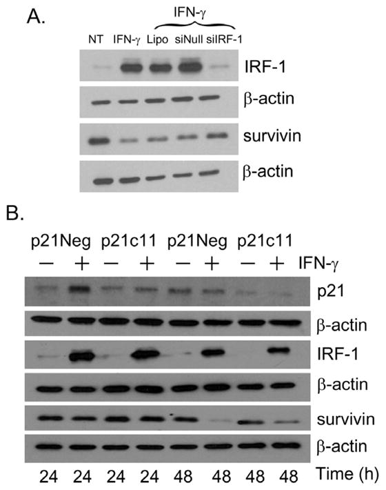
p21-independent suppression of Survivin in human breast cancer cells. (A) MDA-MB-468 cells were either not transfected or transfected with a control siNeg, or siRNA to IRF-1. Untransfected cells were treated with the transfection reagent Lipofectamine (Lipo). 24 h post transfection, the cells were either not treated (NT) or cultured with 1000 U/mL of IFN-γ. 24 h post treatment, cells were harvested and immunoblotting was performed as described in Materials and Methods. (B) p21 psilencer control and p21c11 p21 knockdown MDA-MB-468 clones were either untreated or cultured with 1000 U/mL of IFNγ. Cells were harvested at the 24 and 48 h time points and Western immunoblotting was conducted as has been described in Materials and Methods.
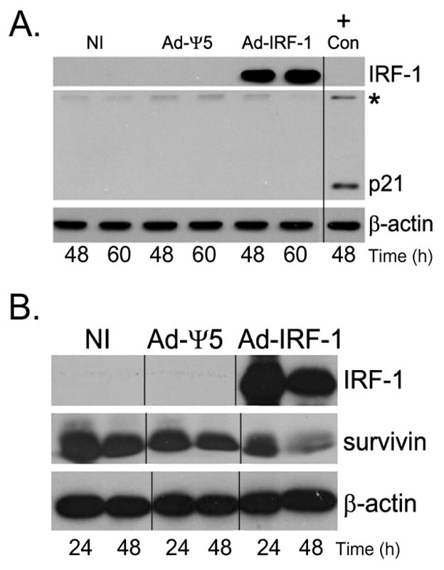
Survivin expression is down-regulated in Ad-IRF-1 infected HCT116 p21−/− human colon cancer cells. (A) HCT 116 p21−/− cells were infected at MOI of 10 and cells were harvested at the indicated time points post infection. IRF-1, p21, and β-actin were assessed by immunoblotting as described in Materials and Methods. On a separate gel run at the same time HCT116 wild type cell lysates were assessed for p21 expression and used as a positive control. * marks a non-specific band that was used to align the separate blots run and stained at the same time. Molecular weight markers were also used for alignment. A p21 band is absent in the HCT116 p21−/− cells regardless of infection. (B) Survivin is decreased by the ectopic IRF-1 expression in HCT116 p21−/− cells. HCT116 p21−/− cells were either uninfected or infected with Ad-Ψ5 or Ad-IRF-1. Cells were harvested at the indicated time points post infection and IRF-1, survivin, and β-actin were assessed by immunoblotting as described in Materials and Methods.
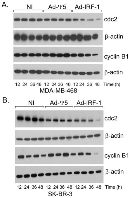
Ectopic expression of IRF-1 results in the down-regulation of cdc-2 and cyclin B1 in human breast cancer cells. (A) The human breast cancer cell line, MDA-MB-468, was either uninfected (NI) or infected with the Ad-Ψ5 or Ad-IRF-1 as has been previously described. Cells were harvested at various time points post infection and Western immunoblotting was conducted as has been described in Materials and Methods. (B) The SK-BR-3 human breast cancer cell line was either uninfected (NI) or infected with the Ad-Ψ5 or Ad-IRF-1 as has been previously described.
References
-
- Ogasawara K, Hida S, Azimi N, Tagaya Y, Sato T, Yokochi-Fukuda T, Waldmann TA, Taniguchi T, Taki S. Requirement for IRF-1 in the microenvironment supporting development of natural killer cells. Nat. 1988;391:700–703. - PubMed
-
- Yim JH, Wu SJ, Casey MJ, Norton JA, Doherty GM. IFN regulatory factor-1 gene transfer into an aggressive, nonimmunogenic sarcoma suppresses the malignant phenotype and enhances immunogenicity in syngeneic mice. J Immunol. 1997;158:1284–1292. - PubMed
-
- Yim JH, Wu SJ, Lowney JK, Vander Velde TL, Doherty GM. Enhancing in vivo tumorigenicity of B16 melanoma by overexpressing interferon regulatory factor-2: resistance to endogenous IFN-gamma. J Interferon Cytokine Res. 1999;19:723–729. - PubMed
-
- Kim PK, Armstrong M, Liu Y, Yan P, Bucher B, Zuckerbraun BS, Gambotto A, Billiar TR, Yim JH. IRF-1 expression induces apoptosis and inhibits tumor growth in mouse mammary cancer cells in vitro and in vivo. Oncogene. 2004;23:1125–1135. - PubMed
Publication types
MeSH terms
Substances
Grants and funding
LinkOut - more resources
Full Text Sources
Molecular Biology Databases
Research Materials
Miscellaneous
