. 2012 Mar;180(3):1145-1158.
doi: 10.1016/j.ajpath.2011.11.006. Epub 2011 Dec 23.

Collagen VI ablation retards brain tumor progression due to deficits in assembly of the vascular basal lamina

Affiliations

Collagen VI ablation retards brain tumor progression due to deficits in assembly of the vascular basal lamina

Weon-Kyoo You et al. Am J Pathol. 2012 Mar.

Abstract

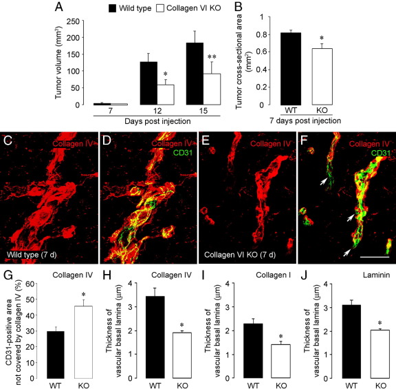
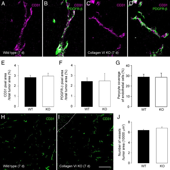
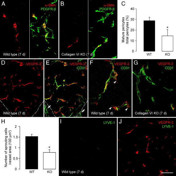
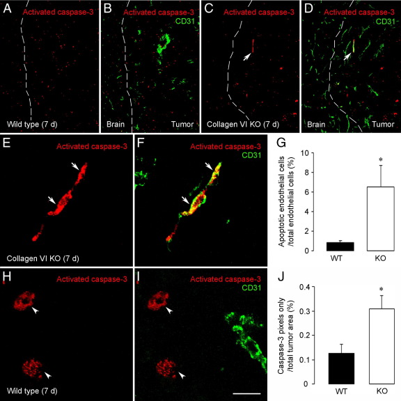
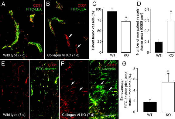
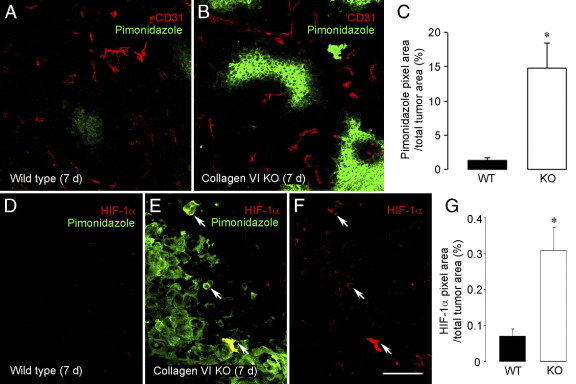
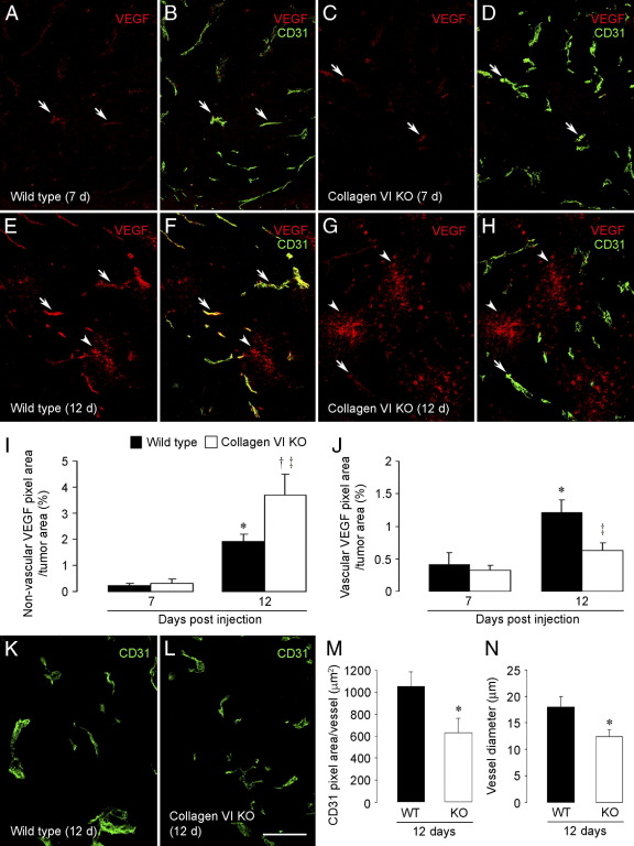
To investigate the importance of the vascular basal lamina in tumor blood vessel morphogenesis and function, we compared vessel development, vessel function, and progression of B16F10 melanoma tumors in the brains of wild-type and collagen VI-null mice. In 7-day tumors in the absence of collagen VI, the width of the vascular basal lamina was reduced twofold. Although the ablation of collagen VI did not alter the abundance of blood vessels, a detailed analysis of the number of either pericytes or endothelial cells (or pericyte coverage of endothelial cells) showed that collagen VI-dependent defects during the assembly of the basal lamina have negative effects on both pericyte maturation and the sprouting and survival of endothelial cells. As a result of these deficits, vessel patency was reduced by 25%, and vessel leakiness was increased threefold, resulting in a 10-fold increase in tumor hypoxia along with a fourfold increase in hypoxia-inducible factor-1α expression. In 12-day collagen VI-null tumors, vascular endothelial growth factor expression was increased throughout the tumor stroma, in contrast to the predominantly vascular pattern of vascular endothelial growth factor expression in wild-type tumors. Vessel size was correspondingly reduced in 12-day collagen VI-null tumors. Overall, these vascular deficits produced a twofold decrease in tumor volume in collagen VI-null mice, confirming that collagen VI-dependent basal lamina assembly is a critical aspect of vessel development.


Figures

Figure 1
Figure 1
Collagen VI ablation retards melanoma progression and reduces basal lamina assembly in tumor vessels. A: After microinjection of B16F10 melanoma cells (104 cells) into the corpus callosum of wild-type and collagen VI-null mice, tumor sizes were measured at 7, 12, and 15 days after injection. At 12 and 15 days, the average tumor size in collagen VI-null mice was twofold smaller than wild-type mice. *P = 0.0362, **P = 0.0483 versus wild type at each time point. B: At 7 days after injection, the average cross-sectional area of tumors in collagen VI-null mice was 80% of that measured in wild-type mice. *P = 0.0222 versus wild type. C–F: To evaluate basal lamina assembly associated with tumor blood vessels, sections of 7-day tumors from wild-type and collagen VI-null mice were double-stained for collagen IV (red, C) and CD31 (green, D). CD31-positive tumor vessels were well covered by the collagen IV matrix in wild-type mice. In contrast, some segments of CD31-positive tumor vessels (F) in collagen VI-null mice were poorly enveloped by the collagen IV-positive basal lamina (E; arrows in F). G: Quantification of collagen IV pixels colocalized with CD31 pixels shows that vascular areas not covered by collagen IV increase from 30% in the wild-type mouse to 46% in the collagen VI-null mouse. *P = 0.0059 versus wild type. H: Moreover, the average width of the collagen IV-positive basal lamina was decreased by 45% in collagen VI-null mice. *P = 0.0047 versus wild type. Immunolabeling for the ECM proteins collagen I (I) and laminin-111 (J) also showed similar reductions in vascular basal lamina width in collagen VI-null mice. *P = 0.0035 (I), *P = 0.03 (J) versus wild type. Scale bars: 20 μm (C–F). KO, knockout; WT, wild type.
Figure 2
Figure 2
Pericyte/endothelial cell dynamics and vessel density in collagen VI-null tumors. To assess pericyte investment of vascular endothelial cells, sections of 7-day tumors from wild-type (A and B) and collagen VI-null (C and D) brains were double-stained for CD31 (purple) and PDGFR-β (green; B and D). E: The density of CD31 pixels is equal in tumors from wild-type mice and collagen VI-null mice. F: This is also true for the density of PDGFR-β pixels. G: In addition, the extent of pericyte coverage of endothelial cells is not affected by collagen VI ablation, as determined by quantifying the percentage of purple pixels that are covered by green pixels in z-stacks of confocal images. CD31 labeling was used to quantify the number of vessels per unit area in both wild-type (H) and collagen VI-null (I) tumors, showing that vascular density is unchanged by collagen VI ablation (J). Dashed line (I) indicates the border between the tumor and normal brain. Scale bars: 40 μm (A–D); 180 μm (H and I). KO, knockout; WT, wild type.
Figure 3
Figure 3
Pericyte maturation and endothelial cell sprouting are reduced by collagen VI ablation. To investigate pericyte maturation in tumor vessels, sections of 7-day tumors from wild-type (A) and collagen VI-null (B) mice were stained for CD31 to localize vessels (not shown) and also were stained for PDGFR-β (green) and α-SMA (red) to identify nascent and mature pericytes, respectively. C: The population of mature pericytes (ie, positive for both pericyte markers) is reduced by 50% in tumors from collagen VI-null mice (C). *P = 0.0426 versus wild type. To evaluate endothelial cell sprouting, 7-day tumor sections from wild-type (D–F) and collagen VI-null (G) mice were double-stained for VEGFR-3 (red) and CD31 (green). Most tumor vessels in wild-type mice were strongly VEGFR-3 immunoreactive, whereas vessels in normal brain were not positive for VEGFR-3 (E, arrows). Dashed lines (D and E) indicate the border between the tumor and normal brain. Typical sprouting endothelial cells are strongly positive for VEGFR-3 and only weakly positive for CD31. These cells were frequently seen in vessels in wild-type tumors (F, arrowheads), but they were significantly less abundant in vessels from collagen VI-null tumors (G). H: Quantification of these cells as a function of total vascular area showed that the number of sprouting endothelial tip cells was reduced by 50% in collagen VI-null mice. *P = 0.0088 versus wild type. Tumor vessels positive for VEGFR-3 staining (J, red) were not costained for LYVE-1 (I and J, green), showing that these are not lymphatic vessels. Scale bars: 60 μm (A, B, F, and G); 120 μm (D, E, I, and J). KO, knockout; WT, wild type.
Figure 4
Figure 4
Increased endothelial cell apoptosis in tumors from collagen VI-null mice. Apoptotic cells identified by staining for activated caspase-3 (red) were scattered throughout 7-day tumors from wild-type mice (A and B), but they were rarely colabeled for CD31 (green). Activated caspase-3-positive cells were more abundant in collagen VI-null mice (C and D) and were sometimes colabeled for CD31 (arrow). Dashed lines (A–D) indicate the tumor/brain border. Collagen VI-null tumors (E and F) contained significant numbers of elongated cells positive for both activated caspase-3 and CD31 (arrows). G: This apoptotic endothelial cell population increased 6.8-fold in tumors from collagen VI-null mice. *P = 0.0469 versus wild type. Scattered round cells positive for activated caspase-3 (H and I) did not express CD31 in wild-type mice (arrowheads). J: The abundance of these cells increased by a factor of 2.4 in collagen VI-null tumors. *P = 0.0332 versus wild type. Scale bars: 120 μm (A–D); 13.3 μm (E, F, H, and I). KO, knockout; WT, wild type.
Figure 5
Figure 5
Reduced vessel patency and increased vessel leakage in collagen VI-null tumors. A–D: Tumor vessel patency was assessed by intravenous FITC-LEA lectin injection (green; 3-minute circulation period) followed by immunohistochemistry for CD31 (red). Most CD31-positive tumor vessels were also stained by FITC-LEA lectin in tumors from wild-type mice (A). In collagen VI-null mice, some CD31-positive tumor vessels were negative for lectin staining (B, arrows), suggestive of nonpatent vessels. C: Quantitative comparisons of FITC-LEA and CD31 labeling indicate that 25% of tumor vessels in collagen VI-null mice are not functionally connected to the circulation. *P = 0.0066 versus wild type. D: When expressed as vessel number per 10,000 μm2 of tumor area, the occurrence of nonpatent vessels is seen to increase threefold in collagen VI-null tumors. *P = 0.0354 versus wild type. E–G: Leakiness of tumor vessels was assessed by intravenous FITC-dextran injection (green, 10-minute circulation period) followed by tissue fixation. Sections were stained for CD31 (red) to define blood vessel boundaries. Most FITC-dextran in tumors from wild-type hosts was contained within the boundaries of tumor blood vessels (E). By comparison, significant amounts of FITC-dextran were found external to CD31-positive vessels in collagen VI-null mice (F, arrows). The extent of FITC-dextran leakage, expressed as the percentage of total tumor area covered by extravascular green pixels, was increased threefold in collagen VI-null mice (G). *P = 0.0395 versus wild type. Scale bars: 40 μm (A and B); 180 μm (E and F). KO, knockout; WT, wild type.
Figure 6
Figure 6
Increased hypoxia and HIF-1α expression in tumors in collagen VI-null mice. A–C: Hypoxia levels in 7-day tumors were determined after intravenous injection of a pimonidazole hypoxia probe (60 mg/kg, 1-hour circulation period). Tumor sections were double-stained for pimonidazole (green) and CD31 (red). In contrast to very small pimonidazole-positive regions in wild-type tumors (A), areas of intratumoral hypoxia were markedly increased in tumors from collagen VI-null mice (B). Intratumoral hypoxia levels, defined as the percentage of total tumor area covered by pimonidazole pixels, were increased 10-fold in collagen VI-null mice (C). *P = 0.0039 versus wild type. D–G: Levels of HIF-1α expression relative to hypoxia were evaluated by immunolabeling for HIF-1α (red) and pimonidazole (green) in sections of 7-day tumors. Neither hypoxia nor HIF-1α was readily apparent in tumors in wild-type mice (D) but was detected in tumors from collagen VI-null mice (E and F). Sites of HIF-1α up-regulation were usually seen in close proximity to areas of hypoxia (E and F, arrows). HIF-1α expression is increased fourfold in tumors from collagen VI-null mice (G). *P = 0.0256 versus wild type. Scale bars: 120 μm (A and B); 40 μm (D–F). KO, knockout; WT, wild type.
Figure 7
Figure 7
Intratumoral VEGF expression and effects on tumor vessels. A–H: VEGF expression (red) in 7-day and 12-day tumors was evaluated by immunofluorescence. At 7 days in both wild-type (A and B) and collagen VI-null (C and D) tumors, very low levels of VEGF (arrows) were detected in association with CD31-positive vessels (green). At 12 days, VEGF levels were much increased in both wild-type (E and F) and collagen VI-null (G and H) tumors. VEGF immunoreactivity in 12-day tumors was distributed in two distinct patterns: closely associated with CD31-positive blood vessels (E–H, arrows) or more randomly dispersed among tumor cells or stroma (E–H, arrowheads). I and J: Quantification of these patterns of VEGF immunofluorescence shows increased nonvascular VEGF (I) and decreased vascular VEGF (J) in collagen VI-null tumors relative to levels seen in wild-type tumors. (I) *P = 0.0005 versus 7-day wild type; P = 0.0025 versus 7-day collagen VI-null; P = 0.0424 versus 12-day wild type. (J) *P = 0.018 versus 7-day wild type; P = 0.0430 versus 12-day wild type. K–N: Immunolabeling for CD31 (green) also allowed assessment of vessel size in 12-day wild-type (K) and collagen VI-null (L) tumors. Vessel size was quantified according to both the CD31-positive area (M) and diameter (N) of each vessel. By both measurements, vessel size was decreased in collagen VI-null tumors. *P = 0.0489 (M), *P = 0.0394 (N) versus wild type. Scale bars: 120 μm (A–H); 180 μm (K and L). KO, knockout; WT, wild type.

Similar articles

Cited by

References

    1. Armulik A., Abramsson A., Betsholtz C. Endothelial/pericyte interactions. Circ Res. 2005;97:512–523. - PubMed
    1. Kalluri R. Basement membranes: structure, assembly and role in tumour angiogenesis. Nat Rev Cancer. 2003;3:422–433. - PubMed
    1. Davis G.E., Senger D.R. Endothelial extracellular matrix: biosynthesis, remodeling, and functions during vascular morphogenesis and neovessel stabilization. Circ Res. 2005;97:1093–1107. - PubMed
    1. Gaengel K., Genové G., Armulik A., Betsholtz C. Endothelial-mural cell signaling in vascular development and angiogenesis. Arterioscler Thromb Vasc Biol. 2009;29:630–638. - PubMed
    1. Huang F.J., You W.K., Bonaldo P., Seyfried T.N., Pasquale E.B., Stallcup W.B. Pericyte deficiencies lead to aberrant tumor vascularizaton in the brain of the NG2 null mouse. Dev Biol. 2010;344:1035–1046. - PMC - PubMed

Publication types

MeSH terms

Substances