Review
. 2012 Feb;63(4):1525-42.
doi: 10.1093/jxb/err394. Epub 2011 Dec 26.

Plant organellar calcium signalling: an emerging field

Affiliations
Review

Plant organellar calcium signalling: an emerging field

Simon Stael et al. J Exp Bot. 2012 Feb.

Abstract

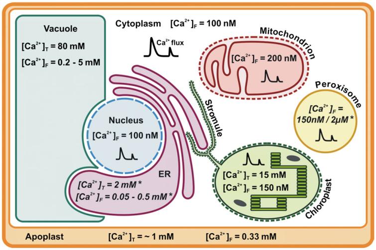
This review provides a comprehensive overview of the established and emerging roles that organelles play in calcium signalling. The function of calcium as a secondary messenger in signal transduction networks is well documented in all eukaryotic organisms, but so far existing reviews have hardly addressed the role of organelles in calcium signalling, except for the nucleus. Therefore, a brief overview on the main calcium stores in plants-the vacuole, the endoplasmic reticulum, and the apoplast-is provided and knowledge on the regulation of calcium concentrations in different cellular compartments is summarized. The main focus of the review will be the calcium handling properties of chloroplasts, mitochondria, and peroxisomes. Recently, it became clear that these organelles not only undergo calcium regulation themselves, but are able to influence the Ca(2+) signalling pathways of the cytoplasm and the entire cell. Furthermore, the relevance of recent discoveries in the animal field for the regulation of organellar calcium signals will be discussed and conclusions will be drawn regarding potential homologous mechanisms in plant cells. Finally, a short overview on bacterial calcium signalling is included to provide some ideas on the question where this typically eukaryotic signalling mechanism could have originated from during evolution.


Figures

Fig. 1
Fig. 1. Summary of organellar Ca2+ concentrations in the plant cell.
Values for reported total ([Ca2+]T) and free resting ([Ca2+]F) Ca2+ concentrations in the different organelles (apoplast, cytoplasm, vacuole, nucleus, ER, chloroplast, mitochondrion, and peroxisome) that were mentioned in the text are depicted here. The values are approximate values and probably vary depending on the tissue or plant species, but nevertheless they provide a general impression of Ca2+ levels across the cell. For ER and peroxisomes, no data on Ca2+ concentration in plants are available. The given values are taken from the animal field and marked with an (*). Calcium fluxes are illustrated by a double peak-shaped symbol (formula image).
Fig. 2
Fig. 2. Overview of proteins involved in calcium signalling in chloroplasts, mitochondria, and peroxisomes of plants.
Calcium and calmodulin (CaM) are highlighted by red boxes. Calcium transporters are depicted in yellow, and calcium/CaM-binding proteins in blue. EF-hand-containing proteins are indicated by the presence of an ‘EF’-box. Proteins of uncertain nature (lack of protein identity or uncertain function) are indicated with a (?). The central ‘Ca2+?’ symbol indicates the possibility of calcium exchange between organelles and the contribution of organelles to the cytoplasmic calcium concentration. Full names for protein abbreviations can be found in the text. Other abbreviations are Calvin (Calvin–Benson cycle), TCA (tricarboxylic acid cycle), IE (inner envelope), OE (outer envelope), IM (inner membrane), OM (outer membrane).
Fig. 3
Fig. 3. Conserved regulators of mitochondrial Ca2+ import between humans and plants.
(A) Alignment of the transmembrane domain of the human mitochondrial calcium uniporter (MCU, gi24308400) with its six homologues from Arabidopsis thaliana displays the two transmembrane helices (TM1 and TM2) connected by a highly conserved loop domain containing the amino acids DIME in human and DVME in Arabidopsis. (B) Overview of targeting and expression of the MCU homologues. The cladogram displays the homology relationship between the Arabidopsis proteins. The blast e-value for each protein relative to the human MCU is written in parentheses after the Arabidopsis gene identifier (AGI). Protein localization was predicted by Aramemnon (Schwacke et al., 2003) and the meta-score is indicated in parentheses. Organ-related RNA expression profiles were examined with the help of the GENEVESTIGATOR browser (Zimmermann et al., 2004) and the presence of expressed sequence tags (ESTs) in the TAIR 10 database (www.arabidopsis.org).

Similar articles

Cited by

References

    1. Aldridge C, Moller SG. The plastid division protein AtMinD1 is a Ca2+-ATPase stimulated by AtMinE1. Journal of Biological Chemistry. 2005;280:31673–31678. - PubMed
    1. Ali R, Ma W, Lemtiri-Chlieh F, Tsaltas D, Leng Q, von Bodman S, Berkowitz GA. Death don’t have no mercy and neither does calcium: Arabidopsis CYCLIC NUCLEOTIDE GATED CHANNEL2 and innate immunity. The Plant Cell. 2007;19:1081–1095. - PMC - PubMed
    1. Ali R, Zielinski RE, Berkowitz GA. Expression of plant cyclic nucleotide-gated cation channels in yeast. Journal of Experimental Botany. 2006;57:125–138. - PubMed
    1. Allen GJ, Muir SR, Sanders D. Release of Ca2+ from individual plant vacuoles by both InsP3 and cyclic ADP-ribose. Science. 1995;268:735–737. - PubMed
    1. Anielska-Mazur A, Bernas T, Gabrys H. In vivo reorganization of the actin cytoskeleton in leaves of Nicotiana tabacum L. transformed with plastin–GFP. Correlation with light-activated hloroplast responses. BMC Plant Biology. 2009;9:64. - PMC - PubMed

Publication types

MeSH terms