. 2012 Jan;139(1):19-30.
doi: 10.1085/jgp.201110717.

The light-sensitive conductance of melanopsin-expressing Joseph and Hesse cells in amphioxus

Affiliations

The light-sensitive conductance of melanopsin-expressing Joseph and Hesse cells in amphioxus

Camila Pulido et al. J Gen Physiol. 2012 Jan.

Abstract

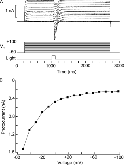
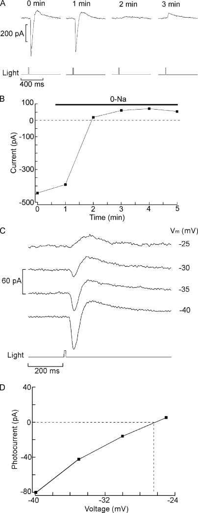
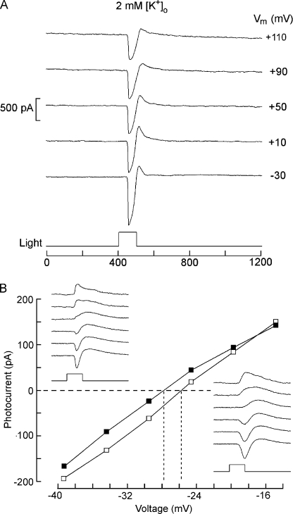
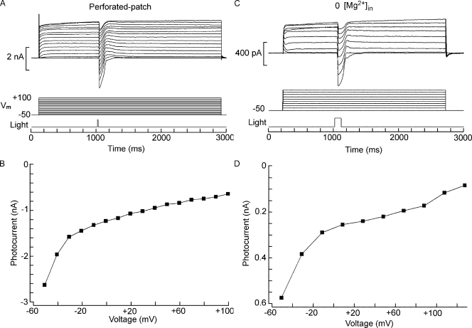
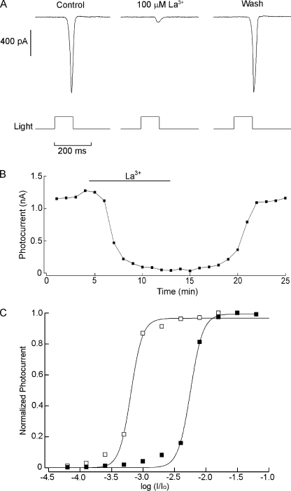
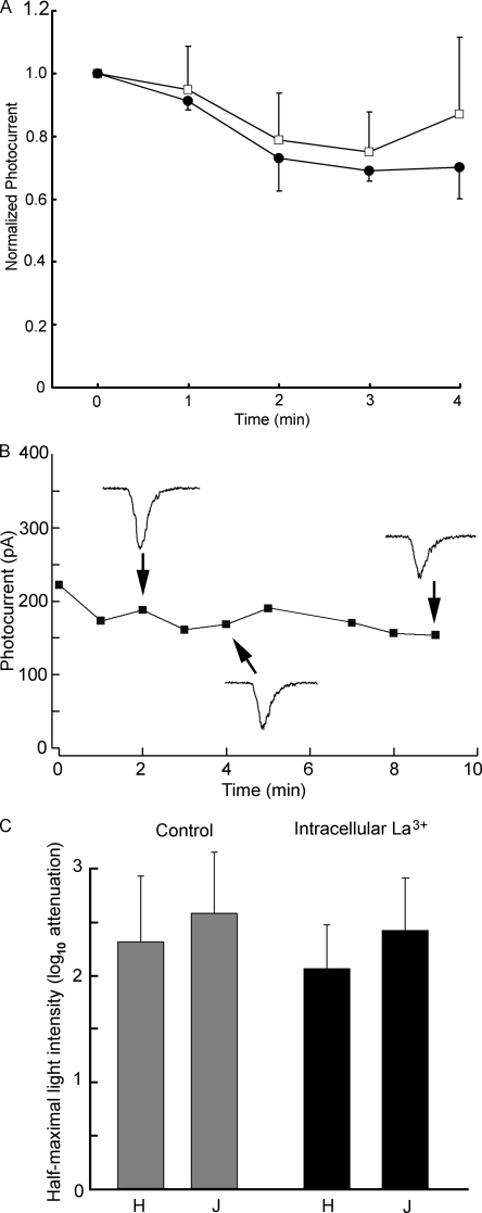
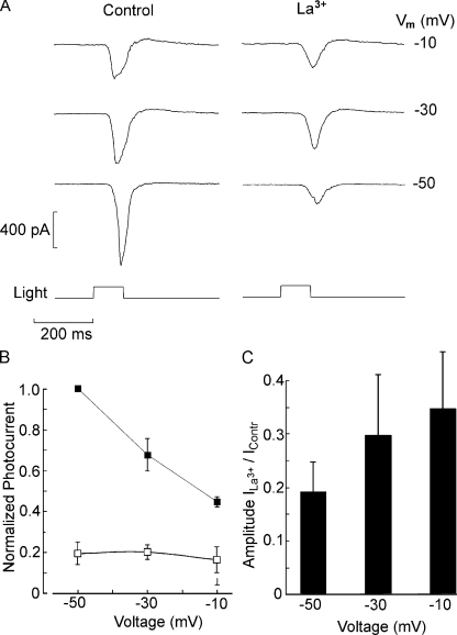
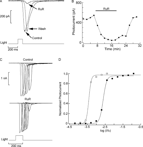
Two types of microvillar photoreceptors in the neural tube of amphioxus, an early chordate, sense light via melanopsin, the same photopigment as in "circadian" light detectors of higher vertebrates. Because in amphioxus melanopsin activates a G(q)/phospholipase C cascade, like phototransduction in arthropods and mollusks, possible commonalities in the photoconductance were investigated. Unlike other microvillar photoreceptors, reversal of the photocurrent can only be attained upon replacement of extracellular Na(+). In addition to Na(+), Ca(2+) is also permeant, as indicated by the fact that (a) in normal ionic conditions the photocurrent remains inward at V(m) > E(Na); (b) in Na-free solution a small residual inward photocurrent persists at V(m) near resting level, provided that Ca is present; and (c) V(rev) exhibits a modest shift with [Ca](o) manipulations. The unusual reversal is accounted for by an uncommonly low permeability of the light-dependent channels to K(+), as [K](o) only marginally affects the photocurrent amplitude and its reversal. Lanthanum and ruthenium red (RuR), two TRP channel antagonists, reversibly suppress the response to photostimulation of moderate intensity; therefore, the melanopsin-initiated cascade may recruit ion channels of the same family as those of rhabdomeric photoreceptors. With brighter lights, blockage declines, so that both La(3+) and RuR induce a right shift in the sensitivity curve without a reduction of its asymptote. Nonetheless, an effect on the transduction cascade, rather than the channels, was ruled out on the basis of the voltage dependency of the blockade and the lack of effects of intracellular application of the same substances. The mechanisms of action of these antagonists thus entail a state-dependent blockade, with a higher affinity for the channel in the closed conformation. Collectively, the results indicate a kinship of the light-sensitive channels of amphioxus with those of invertebrate rhabdomeric visual cells and support the representation of this lineage of photoreceptors among chordates.


Figures

Figure 1.
Figure 1.
Failure to reverse the photocurrent in standard ionic conditions. (A) A Hesse cell was voltage clamped at −50 mV, and depolarizing steps of 2.5-s duration were applied every 60 s, increasing the amplitude at 10-mV increments. At 800 ms after the onset of the voltage stimulus, a 100-ms light flash of constant intensity was delivered (3.8 × 10¹² photons/cm2). Photocurrent size decreased with depolarization but did not reverse polarity, even after stepping membrane voltage up to +100 mV. (B) Peak photocurrent amplitude plotted as a function of membrane voltage, revealing a pronounced inward rectification of the I-V relation.
Figure 2.
Figure 2.
Effects of replacement of extracellular sodium. (A) Joseph cell voltage clamped at 0 mV. After the first light stimulus, the solution was switched to 0 Na/NMDG, causing the photocurrent to become outward. (B) Plot of the photocurrent as a function of time for the full experiment, showing the stable outward light-evoked current in sodium-free solution. Light stimulus, 3.7 × 10¹³ photons/cm2. (C) Photostimulation (4.45 × 10¹³ photons/cm2) applied at different holding voltages to another Joseph cell, showing outwardly directed photocurrent above −30 mV. (D) l-V relation of the photocurrent in sodium-substituted extracellular solution in the same cell as C. The interpolated reversal potential was ≈−26.4 mV.
Figure 3.
Figure 3.
Effects of [K]o manipulations on the amplitude of the light-evoked current. (A) A Hesse cell was voltage clamped and stimulated repetitively; the photocurrent size changed marginally as the extracellular potassium concentration was changed from 10 mM to either 2 or 50 mM. A fixed flash intensity was used throughout (2.8 × 10¹³ photons/cm2), and the holding potential was set at −50 mV. (B) Peak photocurrent amplitude normalized with respect to the value obtained at [K]o = 10 mM, averaged for three Hesse cells tested under the three ionic conditions. Error bars indicate SEM.
Figure 4.
Figure 4.
Effects of extracellular potassium on the reversal of the photocurrent. (A) Increasing the potassium gradient by lowering [K]o to 2 mM in the presence of normal extracellular Na fails to bring about reversal of the light-evoked current when the membrane is depolarized up to +110 mV. Hesse cell: light stimulus, 6.1 × 10¹³ photons/cm2. (B) Effect of reducing [K]o on the reversal potential measured in 0 Na. Sodium in the bath solution was replaced by NMDG, and the light response was examined at either 10 mM [K]o (open symbols; traces of bottom right inset) or 2 mM [K]o (closed symbols; top left inset), as the membrane potential was progressively depolarized at 5-mV increments. The reversal potential of the photocurrent shifted by only 2 mV in the negative direction when external potassium was reduced fivefold.
Figure 5.
Figure 5.
Rectification of the photocurrent. (A and B) The nonlinear conduction of the photocurrent is not an artifact of intracellular perfusion. A Hesse cell was whole cell clamped with the perforated-patch technique using nystatin. After access resistance attained a stable level, the protocol of light stimulation (7.2 × 10¹³ photons/cm2) and membrane depolarization was applied. The resulting light-evoked currents rectified in the inward direction like in control conditions. (C and D) Rectification is not a result of voltage-dependent block by intracellular magnesium ions. Similar experiments were performed with an intracellular solution in which Mg was omitted. The inward rectification was unaffected. Flash intensity: 3.8 × 10¹² photons/cm2).
Figure 6.
Figure 6.
Blockade of the photocurrent by lanthanum. (A) Joseph cell stimulated every minute with light flashes of constant intensity (4.9 × 10¹² photons × s−1cm−2). The panel shows photocurrent traces in control conditions at the peak of the inhibition during bath superfusion with 100 µM and after washing the blocker. (B) The photocurrent amplitude is plotted as a function of time to illustrate the time course of the blockage. (C) Normalized intensity–response curve determined in control conditions and in the presence of 100 µM La3+. Stimulating light was raised at 0.3 log increments. Lanthanum caused a substantial right shift of the curve by approximately one log unit.
Figure 7.
Figure 7.
Effect of intracellular perfusion with La3+. Lanthanum was included in the electrode-filling solution at a concentration of 100 µM, and repetitive photostimulation (one flash/min) started immediately after accessing the cell interior. (A) Normalized average amplitude of the photocurrent in Joseph cells dialyzed with 100 µM lanthanum (closed symbols), compared with control solution (open symbols). No significant differences over time were observed between the two conditions (n = 3 in each group). (B) Similar experiment in a Hesse cell, extending the recording period to 9 min. Raw traces of the photoresponse are shown as insets. (C) Bar graph summarizing the effect of intracellular lanthanum on response sensitivity. The average light intensity required to elicit a response of half-maximal amplitude is plotted for Joseph and Hesse cells tested under the two conditions of intracellular perfusion.
Figure 8.
Figure 8.
Voltage dependence of the block by lanthanum. (A) Photocurrent elicited by a light of approximately half-saturating intensity (2.6 × 10¹¹ photons × s−1cm−2) in a Hesse cell voltage clamped sequentially at −50, −30, and −10 mV in ASW (left) and during exposure to 100 µM La3+ (right). The antagonistic effects of lanthanum diminished as the membrane was depolarized. (B) Pooled data (n = 5, three Hesse and two Joseph cells) showing the average relative amplitude of the photocurrent (normalized by the response in ASW at −50 mV) in control condition (closed squares) and in the presence of lanthanum (open squares). (C) Degree of blockage by lanthanum as a function of membrane potential. The bar graph plots the relative peak amplitude of the photocurrent during superfusion with La3+ versus in ASW.
Figure 9.
Figure 9.
Suppression of the light response by RuR. (A) A Joseph cell voltage clamped at −50 mV was stimulated with flashes of light (2.2 × 10¹¹ photons × s−1cm−2) in ASW during superfusion with 5 µM RuR and after returning to control condition. The photocurrent was reversibly attenuated. (B) Time course of the effect of RuR. The peak amplitude of the light-evoked current is plotted as a function of time for the three phases of the experiment. (C) Intensity series measured in a different Joseph cell in control conditions and in the presence of 5 µM RuR. In the presence of the antagonist, large photocurrents could still be elicited when light intensity was increased. (D) Shift in sensitivity induced by RuR. The normalized peak amplitude of the photocurrent is plotted as a function of light attenuation in the two conditions. A substantial right shift of the stimulus–response curve was induced by RuR. Unattenuated light intensity: 5.1 × 10¹⁴ photons × s−1cm−2.

Similar articles

Cited by

References

    1. Angueyra J.M., Pulido C., Malagón G., Nasi E., Gomez M.P. 2011. Melanopsin-expressing amphioxus photoreceptors transduce light via a phospholipase C signaling cascade. PLoS ONE. In press - PMC - PubMed
    1. Bader C.R., Macleish P.R., Schwartz E.A. 1979. A voltage-clamp study of the light response in solitary rods of the tiger salamander. J. Physiol. 296:1–26 - PMC - PubMed
    1. Bandyopadhyay B.C., Payne R. 2004. Variants of TRP ion channel mRNA present in horseshoe crab ventral eye and brain. J. Neurochem. 91:825–835 10.1111/j.1471-4159.2004.02773.x - DOI - PubMed
    1. Berson D.M. 2003. Strange vision: ganglion cells as circadian photoreceptors. Trends Neurosci. 26:314–320 10.1016/S0166-2236(03)00130-9 - DOI - PubMed
    1. Berson D.M., Dunn F.A., Takao M. 2002. Phototransduction by retinal ganglion cells that set the circadian clock. Science. 295:1070–1073 10.1126/science.1067262 - DOI - PubMed

Publication types

MeSH terms