. 2012 Jan;139(1):83-91.
doi: 10.1085/jgp.201110712.

Light inactivation of water transport and protein-protein interactions of aquaporin-Killer Red chimeras

Affiliations

Light inactivation of water transport and protein-protein interactions of aquaporin-Killer Red chimeras

Florian Baumgart et al. J Gen Physiol. 2012 Jan.

Abstract

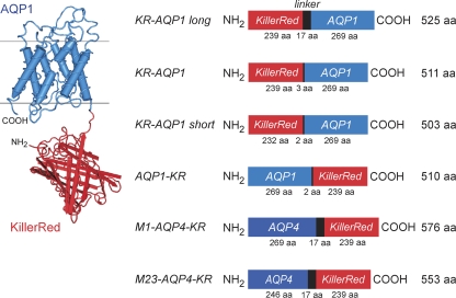
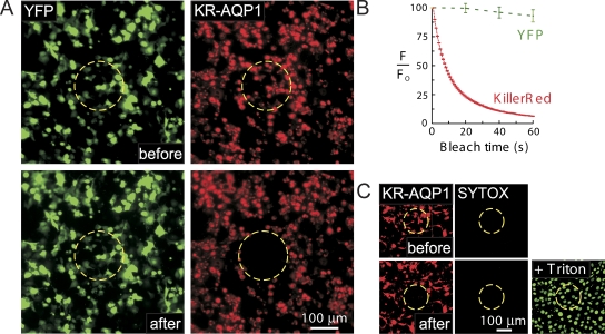
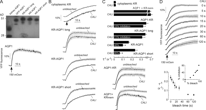
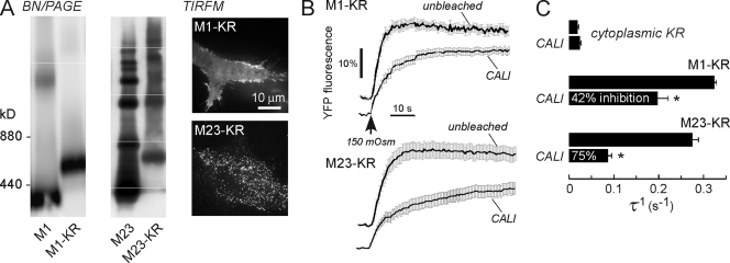
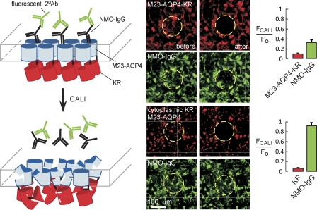
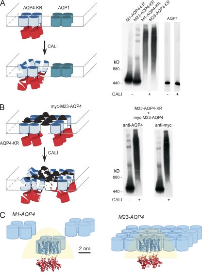
Aquaporins (AQPs) have a broad range of cellular and organ functions; however, nontoxic inhibitors of AQP water transport are not available. Here, we applied chromophore-assisted light inactivation (CALI) to inhibit the water permeability of AQP1, and of two AQP4 isoforms (M1 and M23), one of which (M23) forms aggregates at the cell plasma membrane. Chimeras containing Killer Red (KR) and AQPs were generated with linkers of different lengths. Osmotic water permeability of cells expressing KR/AQP chimeras was measured from osmotic swelling-induced dilution of cytoplasmic chloride, which was detected using a genetically encoded chloride-sensing fluorescent protein. KR-AQP1 red fluorescence was bleached rapidly (~10% per second) by wide-field epifluorescence microscopy. After KR bleaching, KR-AQP1 water permeability was reduced by up to 80% for the chimera with the shortest linker. Remarkably, CALI-induced reduction in AQP4-KR water permeability was approximately twice as efficient for the aggregate-forming M23 isoform; this suggests intermolecular CALI, which was confirmed by native gel electrophoresis on cells coexpressing M23-AQP4-KR and myc-tagged M23-AQP4. CALI also disrupted the interaction of AQP4 with a neuromyelitis optica autoantibody directed against an extracellular epitope on AQP4. CALI thus permits rapid, spatially targeted and irreversible reduction in AQP water permeability and interactions in live cells. Our data also support the utility of CALI to study protein-protein interactions as well as other membrane transporters and receptors.


Figures

Figure 1.
Figure 1.
KR/AQP chimeras. (left) Schematic of a KR/AQP1 chimera with KR at the AQP1 N terminus. (right) Chimeras generated for CALI studies, including three AQP1 N-terminal chimeras of different lengths. Linkers of 17 and 3 amino acids separated KR and full-length AQP1 in KR-AQP1 long and KR-AQP1, respectively. In KR-AQP1 short, the seven C-terminal amino acids of KR were truncated. AQP1-KR and AQP4-KR (M1 and M23) were generated as C-terminal chimeras.
Figure 2.
Figure 2.
Photobleaching of KR-AQP1. (A) FRT cells coexpressing a YFP chloride sensor (green) and KR-AQP1 short (red) were imaged at 20× magnification before (top) and after (bottom) CALI (exposure to red light for 60 s) in the indicated circular regions (circles). (B) Fluorescence of YFP and KR during CALI (error bars indicate standard error [SE], n = 4). (C) Cell viability assayed after CALI (0.3% Triton X-100 as a positive control). Dead cells were stained green (SYTOX).
Figure 3.
Figure 3.
Reduced osmotic water permeability of KR/AQP1 chimeras after CALI. FRT cells were transfected with YFP alone or together with indicated KR/AQP1 chimeras. (A, top) AQP1 immunoblot after SDS/PAGE of cells expressing indicated KR/AQP1 chimeras. Also shown are AQP1 (in kidney lysate) and FRT cells expressing a GFP-AQP1 chimera. Gels that are apart were run separately. (A, bottom) Time course of YFP fluorescence in cells expressing AQP1 (without KR) after rapid reduction in osmolality of the extracellular solution from 300 to 150 mOsm. (B) Osmotic water permeability as in A (bottom) for FRT cells expressing YFP and KR in cytoplasm, KR targeted to the plasma membrane (KRmem), and indicated KR/AQP1 chimeras. (C) Summary of relative osmotic water permeabilities (expressed as reciprocal exponential time constant τ−1) deduced from experiments in B (n = 5–8 cells on three cover glasses, error bars indicate SE; *, P < 0.01). (D) Dependence of AQP1 water permeability inactivation on KR bleaching for KR-AQP1 short. (D, top) Osmotic water permeability in cells exposed to CALI for the indicated times. (D, bottom) Relative water permeability versus CALI bleach time (n = 5–8 cells on three cover glasses, error bars indicate SE; *, P < 0.01). (D, inset) Percentage inhibition of water permeability versus percentage of KR bleach (reduction in fluorescence).
Figure 4.
Figure 4.
CALI efficiency depends on intermolecular topography. FRT cells were transfected with YFP alone or together with M1-AQP4-KR or M23-AQP4-KR. (A, left) AQP4 immunoblot after BN/PAGE. Gels that are apart were run separately. (A, right) TIRFM showing OAP formation by M23-AQP4-KR but not by M1-AQP4-KR. (B) Osmotic water permeability measured by YFP fluorescence, as in Fig. 3 B. (C) Relative osmotic water permeabilities (error bars indicate SE, n = 5–8 cells on three cover glasses; *, P < 0.01).
Figure 5.
Figure 5.
CALI reduces binding of NMO-IgG to an AQP4/KR chimera. (left) Schematic showing CALI of cells expressing M23-AQP4-KR and labeled with NMO-IgG and a secondary Alexa Fluor 488 anti–human antibody. (top middle) CALI (exposure to red light for 60 s) was performed in the indicated circles on cells expressing M23-AQP4-KR and labeled with NMO-IgG and a secondary Alexa Fluor 488 anti–human antibody. (bottom middle) CALI of control cells expressing cytoplasmic KR and unconjugated M23-AQP4. (right) Quantification of KR bleaching and loss of green Alexa Fluor 488 fluorescence after CALI (n = 4, error bars indicate SE; *, P < 0.01).
Figure 6.
Figure 6.
Intermolecular CALI in KR/AQP4 chimeras. (A, left) Schematic of cells expressing KR/AQP4 chimeras together with unconjugated AQP1. (A, right) AQP4 or AQP1 immunoblot after BN/PAGE of lysates from cells expressing M1-AQP4-KR or M23-AQP4-KR, together with unconjugated AQP1, with and without CALI. (B, left) Schematic of cells expressing M23-AQP4-KR together with myc-tagged M23-AQP4. (A, right) AQP4 and myc immunoblot of BN/PAGE of lysates from cells expressing M23-AQP4-KR together with myc-M23-AQP4, with and without CALI. Gels that are apart were run separately. (C) Schematic of distance and clustering-dependent CALI in M1-AQP4 and in OAP-forming M23-AQP4.

Similar articles

Cited by

References

    1. Bulina M.E., Chudakov D.M., Britanova O.V., Yanushevich Y.G., Staroverov D.B., Chepurnykh T.V., Merzlyak E.M., Shkrob M.A., Lukyanov S., Lukyanov K.A. 2006. A genetically encoded photosensitizer. Nat. Biotechnol. 24:95–99 10.1038/nbt1175 - DOI - PubMed
    1. Crane J.M., Verkman A.S. 2008. Long-range nonanomalous diffusion of quantum dot-labeled aquaporin-1 water channels in the cell plasma membrane. Biophys. J. 94:702–713 10.1529/biophysj.107.115121 - DOI - PMC - PubMed
    1. Crane J.M., Verkman A.S. 2009. Determinants of aquaporin-4 assembly in orthogonal arrays revealed by live-cell single-molecule fluorescence imaging. J. Cell Sci. 122:813–821 10.1242/jcs.042341 - DOI - PMC - PubMed
    1. Davies M.J. 2003. Singlet oxygen-mediated damage to proteins and its consequences. Biochem. Biophys. Res. Commun. 305:761–770 10.1016/S0006-291X(03)00817-9 - DOI - PubMed
    1. Del Bene F., Wyart C., Robles E., Tran A., Looger L., Scott E.K., Isacoff E.Y., Baier H. 2010. Filtering of visual information in the tectum by an identified neural circuit. Science. 330:669–673 10.1126/science.1192949 - DOI - PMC - PubMed

Publication types

MeSH terms