. 2011 Nov;6(6):835-848.
doi: 10.1586/eem.11.70.

Transmembrane mucins as novel therapeutic targets

Affiliations

Transmembrane mucins as novel therapeutic targets

Pamela E Constantinou et al. Expert Rev Endocrinol Metab. 2011 Nov.

Abstract

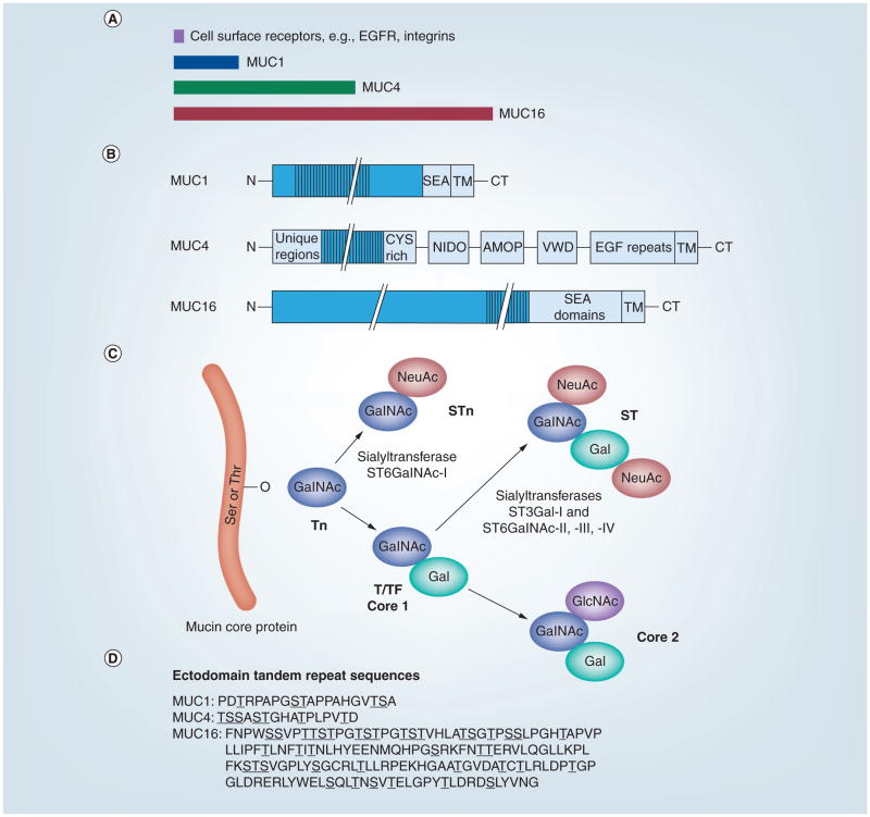
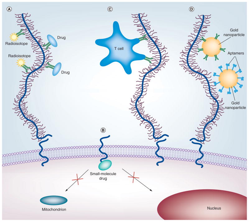
Membrane-tethered mucin glycoproteins are abundantly expressed at the apical surfaces of simple epithelia, where they play important roles in lubricating and protecting tissues from pathogens and enzymatic attack. Notable examples of these mucins are MUC1, MUC4 and MUC16 (also known as cancer antigen 125). In adenocarcinomas, apical mucin restriction is lost and overall expression is often highly increased. High-level mucin expression protects tumors from killing by the host immune system, as well as by chemotherapeutic agents, and affords protection from apoptosis. Mucin expression can increase as the result of gene duplication and/or in response to hormones, cytokines and growth factors prevalent in the tumor milieu. Rises in the normally low levels of mucin fragments in serum have been used as markers of disease, such as tumor burden, for many years. Currently, several approaches are being examined that target mucins for immunization or nanomedicine using mucin-specific antibodies.


Figures

Figure 1
Figure 1. Membrane-bound mucins
(A) The sizes of typical cell surface receptors, such as intergrins and EGFR, relative to those of MUC1, MUC4 and MUC16 are shown. (B) Domain organization of MUC1, MUC4 and MUC16. (C) Cancer-associated antigens (Tn, STn and T/TF) result from aberrant O-glycosylation on the ectodomains of MUC1, 4 and 16. O-linked glycosylation is initiated by adding GalNAc to the protein backbone, resulting in the Tn antigen. NeuAc of the GalNAc moiety results in the formation of the STn antigen. Alternatively, core 1 structures, T and TF, are synthesized by the addition of Gal to the Tn antigen. Non-cancer-associated antigens are formed from either the sialylation of the core 1 structures, resulting in the mono- or disialyl-T antigen, or the continued branching/elongation by adding GlcNAc to form the core 2 structures. Further elongation of the core 2 structure forms more complex peripheral antigens, such as the variations of Lewis and Sialyl–Lewis. (D) O-glycosylation occurs on either a Ser or Thr residue within the tandem repeat sequences of the mucin ectodomains. The consensus sequences for the tandem repeats in human MUC1, MUC4 and MUC16 are shown using the single letter amino acid code. Serine and threonine residues are underlined. Note the large number of proline residues in these motifs. AMOP: Adhesion-associated domain; CT: Cytoplasmic tail; CYS rich: Cysteine-rich domain; EGF repeats: EGF-like repeat region; EGFR: EGF receptor; Gal: Galactose; GalNAc: N-acetylgalactosamine; GlcNAc: N-acetylglucosamine; MUC: Mucin; NeuAc: Sialylation; NIDO: Nidogen-like domain; SEA: Sea urchin sperm receptor enterokinase-agrin domain; Ser: Serine; ST: Disialyl-T; STn: Sialyl-Tn; T/TF: Thomsen–Friedenreich antigen; Thr: Threonine; TM: Transmembrane domain; Tn: Precursor to T antigen; VWD: von Willebrand factor type D domain.
Figure 2
Figure 2. Transmembrane mucins as therapeutic targets
(A) Antibody-based therapies include radioisotopes (yellow star shapes) and drugs (blue ovals) conjugated to antibodies (green Y-shapes) recognizing the ectodomains of mucins. (B) Peptides and small-molecule drugs (turquoise oval) can block the active cytoplasmic domain of MUC1, thus preventing its localization to the mitochondria or nucleus. (C) Immunotherapies designed to stimulate the body’s immune response and induce T cells (blue amoeboid shape) that target mucins. (D) Targeted nanotherapies use antibodies or aptamers (blue hook shapes) that recognize the ectodomains of mucins to deliver gold nanoparticles (large orange circles) or quantum dots. For details, see text.

Similar articles

Cited by

References

    1. Kufe DW. Mucins in cancer: function, prognosis and therapy. Nat Rev Cancer. 2009;9(12):874–885. - PMC - PubMed
    1. Hollingsworth MA, Swanson BJ. Mucins in cancer: protection and control of the cell surface. Nat Rev Cancer. 2004;4(1):45–60. - PubMed
    1. Choudhury A, Moniaux N, Ringel J, et al. Alternate splicing at the 3′-end of the human pancreatic tumor-associated mucin MUC4 cDNA. Teratog Carcinog Mutagen. 2001;21(1):83–96. - PubMed
    1. Wreschner DH, Hareuveni M, Tsarfaty I, et al. Human epithelial tumor antigen cDNA sequences. Differential splicing may generate multiple protein forms. Eur J Biochem. 1990;189(3):463–473. - PubMed
    1. Ligtenberg MJ, Kruijshaar L, Buijs F, van Meijer M, Litvinov SV, Hilkens J. Cell-associated episialin is a complex containing two proteins derived from a common precursor. J Biol Chem. 1992;267(9):6171–6177. - PubMed

Websites

    1. National Institutes of Health Registry of Clinical Trials. www.clinicaltrials.gov.
    1. Press release: Therion Reports Results Of Phase 3 PANVAC-VF Trial And Announces Plans For Company Sale. www.medicalnewstoday.com/releases/46137.php.