Review
. 2012 Apr;1817(4):672-9.
doi: 10.1016/j.bbabio.2011.12.005. Epub 2011 Dec 16.

Kinetic studies of the reactions of O(2) and NO with reduced Thermus thermophilus ba(3) and bovine aa(3) using photolabile carriers

Affiliations
Review

Kinetic studies of the reactions of O(2) and NO with reduced Thermus thermophilus ba(3) and bovine aa(3) using photolabile carriers

Olöf Einarsdóttir et al. Biochim Biophys Acta. 2012 Apr.

Abstract

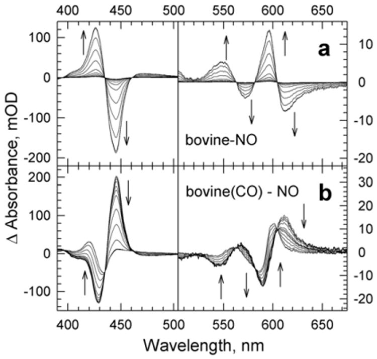
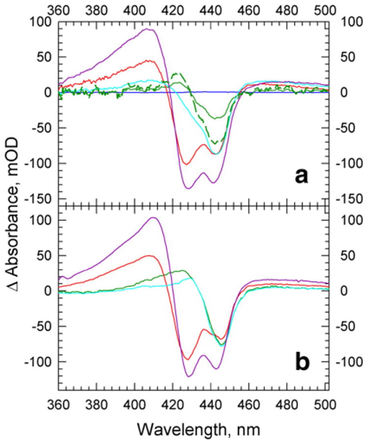
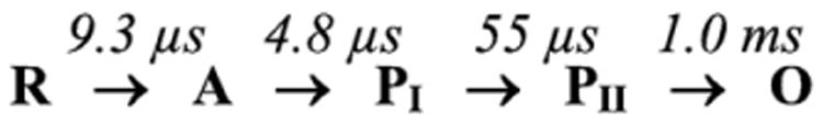
The reactions of molecular oxygen (O(2)) and nitric oxide (NO) with reduced Thermus thermophilus (Tt) ba(3) and bovine heart aa(3) were investigated by time-resolved optical absorption spectroscopy to establish possible relationships between the structural diversity of these enzymes and their reaction dynamics. To determine whether the photodissociated carbon monoxide (CO) in the CO flow-flash experiment affects the ligand binding dynamics, we monitored the reactions in the absence and presence of CO using photolabile O(2) and NO complexes. The binding of O(2)/NO to reduced ba(3) in the absence of CO occurs with a second-order rate constant of 1×10(9)M(-1)s(-1). This rate is 10-times faster than for the mammalian enzyme, and which is attributed to structural differences in the ligand channels of the two enzymes. Moreover, the O(2)/NO binding in ba(3) is 10-times slower in the presence of the photodissociated CO while the rates are the same for the bovine enzyme. This indicates that the photodissociated CO directly or indirectly impedes O(2) and NO access to the active site in Tt ba(3), and that traditional CO flow-flash experiments do not accurately reflect the O(2) and NO binding kinetics in ba(3). We suggest that in ba(3) the binding of O(2) (NO) to heme a(3)(2+) causes rapid dissociation of CO from Cu(B)(+) through steric or electronic effects or, alternatively, that the photodissociated CO does not bind to Cu(B)(+). These findings indicate that structural differences between Tt ba(3) and the bovine aa(3) enzyme are tightly linked to mechanistic differences in the functions of these enzymes. This article is part of a Special Issue entitled: Respiratory Oxidases.


Figures

Fig. 1
Fig. 1
Time-resolved optical absorption difference spectra (post-minus pre-photolysis) of the reaction of the fully reduced Tt ba3 with photoproduced NO in the absence of CO (panel a) and the presence of CO (panel b). The spectra were obtained by subtracting the spectral contribution of the photolyzed NO complex, determined in a separate experiment. The spectra (SVD-filtered) were recorded at 12–16 delay times, equally spaced on a logarithmic scale, between 200 ns–10 ms in the absence of CO and 500 ns–50 ms in the presence of CO. The arrows represent the direction of the absorbance change with time. Conditions: the effective enzyme concentration, 1.2 μM; 0.1 M HEPES (pH 7.5); 0.1% DM; effective NO concentration, 60 μM (panel a) and70 μM (panel b); optical path, 0.5 cm. TheCOconcentration was 0.5 mMafter mixing.
Fig. 2
Fig. 2
Time-resolved optical absorption difference spectra for the reaction of the fully reduced bovine aa3 with photoproduced NO in the absence of CO (panel a; 100 ns–10 ms) and the presence of CO (panel b; 200 ns–10 ms) (see Fig. 1 for details). Conditions: the effective enzyme concentration, 5.2 μM; 50 mM Na-phosphate (pH 7.5); effective NO concentration: 87 μM (a) and 105 μM (b). The CO concentration was 0.5 mM after mixing.
Fig. 3
Fig. 3
Comparison of the transient absorbance changes taking place at 444 nm during the reaction of fully reduced Tt ba3 (panel a) and bovine aa3 (panel b) with photoproduced NO in the presence of CO (filled circles and triangles) and in the absence of CO (open circles and triangles). The kinetic traces in panels a and b are from the time-resolved data in Figs. 1 and 2, respectively, and are normalized to the total absorbance change. The solid lines represent the absorbance traces at 444 nm, calculated on the basis of the global exponential fits (see text for details).
Fig. 4
Fig. 4
The spectral amplitudes (b-spectra; solid curves) and lifetimes from a global-exponential fit to the time-resolved data recorded during the reaction of NO with reduced Tt ba3 in the absence (a) and presence (b) of CO. The time-independent bo-spectra (dashed curves) represent the difference spectra extrapolated to infinite time, namely, the difference between the spectra of the NO-bound and reduced enzymes (a), and the NO-bound and CO-bound enzymes (b).
Fig. 5
Fig. 5
Time-resolved optical absorption difference spectra (post-minus pre-photolysis) recorded during the reaction of dioxygen with the fully reduced Tt ba3 (panel a) and bovine enzyme (panel b) in the absence of CO. The spectra are those obtained after subtracting the spectral contribution of the photolyzed O2 complex, determined in a separate experiment. The spectra (SVD-filtered) were recorded at 15–17 delay times, equally spaced on a logarithmic scale, between 500 ns–20 ms (panel a) and 200 ns– 50 ms (panel b). The arrows represent the direction of the absorbance change with time. Conditions: ba3: 0.1 M HEPES (pH 7.5); 0.1% DM; effective enzyme concentration, 2.6 μM; bovine enzyme: 50 m N NaPi (pH 7.5) effective enzyme concentration, 4.3 μM; optical path, 0.5 cm.
Fig. 6
Fig. 6
(Panel a) (Solid lines) The experimental intermediate spectra (referenced versus the reduced enzyme) for the reaction of reduced Tt ba3 with O2 in the absence of CO. The spectra were extracted on the basis of the fast–slow mechanism in Scheme 2: compound A (green), PI (red), PII (cyan) and O (magenta). The spectrum of Int 2 extracted using the fast–slow Scheme 2 has the shape of compound A of the bovine heart oxidase but significantly lower amplitude. (Dashed, green curve) The spectrum of compound A extracted using the slow–fast mechanism in Scheme 3, in which the 9.3 μs process is followed by the 4.8 μs step. (Panel b) The model spectra of the proposed intermediates, compoundA(green), PI (red),PII (cyan) and O (magenta), were generated basedonthelinear combinations of the ground-state spectra of ba3; the difference spectrum of compound A is thatof the bovine enzyme. The spectrain panel a are normalized to the enzyme concentration in panel b.
Scheme 1
Scheme 1
The conventional sequential unidirectional mechanism for the reduction of dioxygen to water.
Scheme 2
Scheme 2
A unidirectional “fast-slow” sequential mechanism of O2 reduction for ba3 in the absence of CO.
Scheme 3
Scheme 3
The “slow-fast” sequential mechanism for the reaction of ba3 with photo-produced O2 in the absence of CO.

Similar articles

Cited by

References

    1. Brzezinski P, Larsson G. Redox-driven proton pumping by heme–copper oxidases. Biochim Biophys Acta. 2003;1605:1–13. - PubMed
    1. Ferguson-Miller S, Babcock GT. Heme/copper terminal oxidases. Chem Rev. 1996;96:2889–2907. - PubMed
    1. Kaila VR, Verkhovsky MI, Wikström M. Proton-coupled electron transfer in cytochrome oxidase. Chem Rev. 2010;110:7062–7081. - PubMed
    1. Einarsdóttir Ó. Fast reactions of cytochrome oxidase. Biochim Biophys Acta. 1995;1229:129–147. - PubMed
    1. Wikström MKF. Proton pump coupled to cytochrome c oxidase in mitochondria. Nature. 1977;266:271–273. - PubMed

Publication types

MeSH terms

LinkOut - more resources