. 2012 Mar;234(1):50-61.
doi: 10.1016/j.expneurol.2011.12.016. Epub 2011 Dec 19.

Effects of aging on blood brain barrier and matrix metalloproteases following controlled cortical impact in mice

Affiliations

Effects of aging on blood brain barrier and matrix metalloproteases following controlled cortical impact in mice

Phil Lee et al. Exp Neurol. 2012 Mar.

Abstract

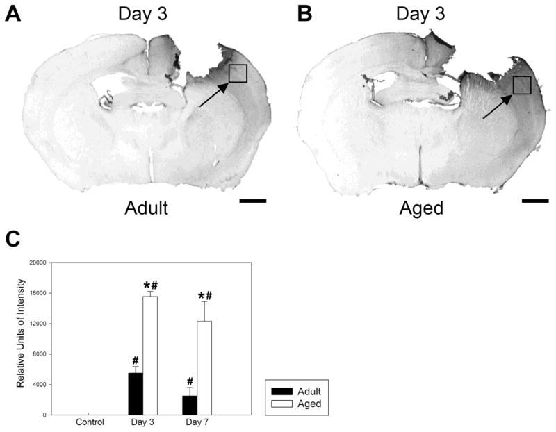
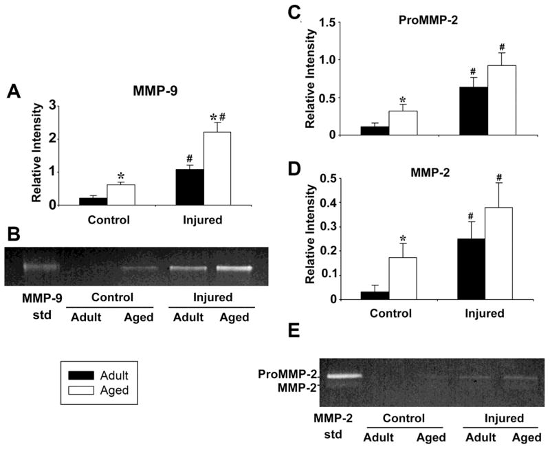
Aging alters the ability of the brain to respond to injury. One of the major differences between the adult and aged brain is that comparable injuries lead to greater blood brain barrier disruption in the aged brain. The goals of these studies were to quantify the effects of age on BBB permeability using high field strength MRI T1 mapping and to determine whether activation of matrix metalloproteases, their inhibitors, or expression of blood brain barrier structural proteins, occludin, zonnula occludins-1 (ZO-1) and claudin-5 were altered following injury to the aged C57/BL6 mouse brain. T1 mapping studies revealed greater blood brain barrier permeability in the aged (21-24 months old) brain than in the adult (4-6 months old) following controlled cortical impact. The increased blood brain barrier permeability in the pericontusional region was confirmed with IgG immunohistochemistry. MMP-9 activity was increased following controlled cortical impact in the aged brain, and this was accompanied by increased MMP-9 gene expression. MMP-2 activity was higher in the uninjured aged brain than in the adult brain. Occludin and ZO-1 mRNA levels were unchanged following injury in either age group, but claudin-5 mRNA levels were lower in the aged than the adult brain following injury. These results demonstrate quantitative increases in blood brain barrier permeability in the aged brain following injury that are accompanied by increased MMP-9 activation and decreased blood brain barrier repair responses.


Figures

Figure 1
Figure 1. Time course of T1 contrast enhancement of a mouse brain following i.p. Gd-DTPA administration
A. T1 maps were acquired at before (pre), 1, 2, 3, and 4 hr after Gd-DTPA (0.2 mmol/kg) administration. The areas of interest where the time course of R1 (=1/T1) were obtained are marked as squares 1 (core) and 3 (peri-contusional area). The vertical gray scale bar indicates the range of T1 values. B. Time courses of R1 of the core (open circle) and peri-contusional area (filled square). Error bars indicate +/−SE.
Figure 2
Figure 2. T2 and T1 mapping of gadolinium enhancement in adult and aged mouse brain after controlled cortical impact
A. T2-weighted image (left column) and corresponding T1 maps acquired before (middle column) and 2 hr after (right column) Gd-DTPA administration in an adult mouse. Images in the top row were acquired at day 3 and images at the bottom row were acquired at day 14. Three areas of interest marked by 1 (core), 2 (contra-contusional area) and 3 (peri-contusional area) are shown in the T2-weighted image at D14 (bottom left). The gray scale bars indicate the range of T1 values between 0 to 2.3 s. B. R1 changes at the core of contusional area (1) after Gd-DTPA administration in adult (black bars) and aged mice (white bars) acquired at days 3, 7 and 14. Error bars indicate +/− SE. C. R1 changes at the peri-contusional area (3) after Gd-DTPA administration in adult (black bars) and aged mice (white bars). D. R1 changes at the contra-contusional area after Gd-DTPA. Error bars indicate +/− SE. N=6 adult, 6 aged mice.
Figure 3
Figure 3. Immunohistochemistry of extravasated IgG reveals greater blood brain barrier disruption following controlled cortical impact in aged than in adult mice
A. IgG staining in an adult brain, 3 days post CCI. B. IgG staining in an aged mouse brain, 3 days post CCI. In both cases, the boxes indicated by arrows outline the regions in which image intensity was measured. C. Quantitation of staining intensity in the region shown in A and B. Both the adult and aged mice showed increased IgG staining in the outlined region at 3 and 7 days (p<0.05). In addition, the aged mice had significantly more IgG staining at day 3 after injury (p-value < 0.001) and day 7 after injury (p < 0.05) than adult injured mice. Differences from uninjured values are indicated by #, while differences between adult and aged values are indicated by *. There were no significant IgG staining differences in this region of the cortex between aged and adult non-injured control mice. Error bars indicate +/−SE. N= 5 adult control, 3 aged control, 6 adult injured day 3, 6 aged injured day 3, 4 adult injured day 7, 4 aged injured day 6. Scale bar = 1mm.
Figure 4
Figure 4. MMP activity after controlled cortical impact is higher in the aged than in the adult mouse brain
Quantitation of zymographic assay of (A) MMP-9 activity in injured cortex 3 days after CCI in adult and aged mice. B. Zymogram of MMP-9 activity including standard (MMP-9 std). C. Quantitation of zymographic assay of pro-MMP-2 activity in injured cortex 3 days after CCI in adult and aged mice. D. Quantitation of zymographic assay of MMP-2 activity in injured cortex 3 days after CCI in adult and aged mice. E. Image of zymogram of proMMP-2 and active MMP-2 including standards (MMP-2 std). The increase in the MMP-9 baseline activity and the difference in injury response between adult and aged mice are both statistically significant, while the difference in baseline MMP-2 activity between adult and aged mice is significant. *= p<.05 adult vs. aged, #= p<0.05 injured vs. control. N=3 adult controls, 3 aged controls, 3 adult injured, 3 aged injured. Error bars indicate +/− SE
Figure 5
Figure 5. Assessment of gene expression of MMP, TIMP, and blood brain barrier structural proteins following controlled cortical impact in the adult and aged brain
Gene expression was measured in uninjured cortex (D0) and at 1,2,3,7 and 14 days post CCI. A. MMP-9, B. MMP-2, C. TIMP-1, D. TIMP-2, E. occludin, F. ZO-1, G. Claudin-5.. All measurements are relative to GAPDH levels in the same sample. D0=uninjured control mice. Y axes are relative expression. *= p<.05 comparing adult vs. aged, #= p<0.05 comparing injured vs. control using two independent sample Wilcoxon rank sum tests. N= Uninjured: 7 adult, 7 aged; D1: 6 adult, 6 aged; D2: 6 adult, 6 aged; D3: 7 adult, 7 aged; D7: 6 adult; 6 aged; D14: 6 adult, 6 aged. Error bars indicate +/− SE

Similar articles

Cited by

References

    1. Adams DD, Lucas WO, Williams BG, Berkeley BB, Turner KW, Schofield JC. A mouse genetic locus with death clock and life clock features. Mech Ageing Dev. 2001;122:173–89. - PubMed
    1. Anderson J, Sandhir R, Hamilton ES, Berman NE. Impaired expression of neuroprotective molecules in the HIF-1alpha pathway following traumatic brain injury in aged mice. J Neurotrauma. 2009;26:1557–66. - PMC - PubMed
    1. Asahi M, Wang X, Mori T, Sumii T, Jung JC, Moskowitz MA, Fini ME, Lo EH. Effects of matrix metalloproteinase-9 gene knock-out on the proteolysis of blood-brain barrier and white matter components after cerebral ischemia. J Neurosci. 2001;21:7724–32. - PMC - PubMed
    1. Berman NE, Marcario JK, Yong C, Raghavan R, Raymond LA, Joag SV, Narayan O, Cheney PD. Microglial activation and neurological symptoms in the SIV model of NeuroAIDS: association of MHC-II and MMP-9 expression with behavioral deficits and evoked potential changes. Neurobiol Dis. 1999;6:486–98. - PubMed
    1. Bilgen M. A new device for experimental modeling of central nervous system injuries. Neurorehabil Neural Repair. 2005;19:219–26. - PubMed

Publication types

MeSH terms