. 2012 Mar;234(1):85-94.
doi: 10.1016/j.expneurol.2011.12.012. Epub 2011 Dec 20.

Traumatic brain injury and trichloroethylene exposure interact and produce functional, histological, and mitochondrial deficits

Affiliations

Traumatic brain injury and trichloroethylene exposure interact and produce functional, histological, and mitochondrial deficits

Andrew Sauerbeck et al. Exp Neurol. 2012 Mar.

Abstract

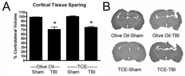
Mitochondria play a pivotal role in the development of pathology associated with Parkinson's disease (PD), traumatic brain injury (TBI), and following exposure to the environmental toxin trichloroethylene (TCE). Evidence from humans indicates that both TBI and TCE can play a role in the development of PD and that each of these insults result in significant mitochondrial dysfunction. In the current studies we hypothesized that exposure to both TCE and TBI would result in increased pathology associated with PD. To test this hypothesis, 16 week old male Fischer 344 rats were administered TCE for either one or two weeks by oral gavage. Following exposure to TCE, rats were subjected to either a sham, mild (1.0mm), or moderate (2.0mm) controlled cortical impact TBI. Given the strong connection between mitochondrial function and PD, TBI, and TCE, tissue from the striatum and substantia nigra were analyzed 6h after the TBI. Neither TCE exposure, TBI, nor the combination of the two insults resulted in mitochondrial deficits at 6h post-TBI in the substantia nigra. Unlike the substantia nigra, the striatum exhibited significant mitochondrial dysfunction. Exposure to TCE alone for two weeks resulted in approximately a 75% reduction in mitochondrial function (p<0.05) in the striatum whereas TBI alone resulted in approximately a 30% reduction in striatal mitochondrial function. Following 1 week exposure to TCE followed by TBI, there was a significant reduction (50%) in mitochondrial function (p<0.05) which required the presence of both insults. Beginning 12 days after the injury significant motor impairment was observed with Rotarod testing. Animals exposed to TCE and a moderate TBI exhibited performance which was approximately 50% of controls (p<0.01). Cylinder testing revealed that at 30 days post-injury animals exposed to TCE and a moderate TBI also had about a 34% reduction in the usage of the contralateral fore paw and this impairment was significantly worse than both control animals and animals exposed to TCE and a mild TBI (p<0.05). At 30 days post-injury there was a 13-17% reduction in the number of tyrosine hydroxylase (TH) positive neurons in the substantia nigra (p<0.05), which was the result of protein loss and not cell death. Loss of TH positive neurons did not result in changes in striatal TH fiber density or levels of the dopamine transporter or type-2 dopamine receptor. Additionally, exposure to TCE prior to the TBI did not increase the loss of cortical tissue, indicating regional specificity for TCE induced deficits. These studies provide further evidence for the connection between TCE, TBI, and PD and lend support to the concept that PD develops from a multifactorial injury scenario.


Figures

Figure 1
Figure 1. 2 week exposure to TCE and TBI results in decreases in mitochondrial bioenergetics in the striatum
Animals were exposed to TCE for 2 weeks and then subjected to a moderate TBI, and mitochondria were isolated from the striatum and substantia nigra six hours post-injury. A) Analysis of bioenergetic function in the substantia nigra revealed no significant difference between any of the treatment groups. B) Exposure to TCE for 2-weeks produced significant reductions of approximately 75% in Complex I dependent basal and maximal rates of mitochondrial oxygen consumption. The TBI alone resulted in a trend towards reduced mitochondrial function in the striatum, however, this decrease did not reach significance. *=p<0.05, n=5-6; 2-Way ANOVA with Bonferroni post-test; Mean±SEM
Figure 2
Figure 2. The combined exposure of TCE for 1 week with a moderate TBI results in impaired in mitochondrial bioenergetics in the striatum
Exposure to either TCE for 1 week or a moderate TBI did not result in significant mitochondrial dysfunction in the striatum. When the two separate insults were combined together there was a significant reduction in the Complex I driven rate of mitochondrial oxygen consumption. The dual injury paradigm resulted in synergistic mitochondrial deficits, which resulted in approximately a 50% reduction in mitochondrial function. *=p<0.05, n=3-4; 2-Way ANOVA with Bonferroni post-test; Mean±SEM
Figure 3
Figure 3. Exposure to TCE and a moderate TBI synergistically interact resulting in functional impairment
Exposure to both TCE for 2 weeks and a moderate controlled cortical impact TBI result in significant motor impairment. A) Twelve days after the TBI animals exhibited significant motor impairment, as exhibited by a reduced ability to stay on a rotating rod. Animals exposed to TCE and a moderate TBI have approximately a 50% reduction in the length time able to stay on the Rotarod.B) Thirty days after the TBI, forepaw usage was assessed using the cylinder test and contralateral forepaw usage decreased to 34%. These results demonstrate the necessity for both insults to be present before functional impairment occurs. #=p<0.01, *=p<0.05; n=6; 2-Way ANOVA with Bonferroni post-test; Mean±SEM
Figure 4
Figure 4. Expose to TCE and TBI interact and result in TH-positive neuron loss in the Substantia Nigra
Exposure to both TCE for 2 weeks and a mild or moderate controlled cortical impact TBI resulted in significant decreases in the number of TH-positive neurons in the substantia nigra at 30 days post-TBI. A) The dual injury consisting of TCE exposure and a TBI brain injury resulted in a 13-17% loss of dopamine neurons in the substantia nigra. B) Quantification of Nissl cells in the substantia nigra did not reveal any measurable amount of cell loss, indicating the loss of TH-positive neurons is due to a loss of the TH protein and not the loss of cells. C) Representative images of the substantia nigra stained for TH. *=p<0.05; n=6; 2-Way ANOVA with Bonferroni post-test; Mean±SEM
Figure 5
Figure 5. Exposure to TCE, TBI, or the dual injury does not lead to striatal changes in TH-fibers, DAT, and D2R
Neither exposure to TCE for 2 weeks, a mild or moderate TBI, or any combination of the insults resulted in histological deficits in the striatum. A) The amount of area in the striatum immunoreactive for TH was quantified and no reduction in TH-positive fibers was measured. B) Representative images of the striatum stained for TH. C,D) Western blot analysis for levels of the dopamine transporter and type-2 dopamine receptor revealed no changes in either protein. N=6; 2-Way ANOVA; Mean±SEM. Data are expressed as a ratio to level of β-actin.
Figure 6
Figure 6. Exposure to TCE prior to a moderate TBI does not increase cortical tissue loss
Animals that were exposed to TCE for 2 weeks prior to a moderate TBI did not exhibit an increase in the amount of cortical tissue loss. A) With this model a moderate TBI resulted in approximately a 30% loss of cortical tissue at 30 days post-injury. Exposure to TCE had no effect on the loss of cortical tissue. B) Representative images of Nissl stained cortex 30 days post-injury. *=p<0.0001; n=6; 2-Way ANOVA with Bonferroni post-test; Mean±SEM

Similar articles

Cited by

References

    1. Afonso-Oramas D, Cruz-Muros I, Alvarez de la Rosa D, Abreu P, Giraldez T, Castro-Hernandez J, Salas-Hernandez J, Lanciego JL, Rodriguez M, Gonzalez-Hernandez T. (Dopamine transporter glycosylation correlates with the vulnerability of midbrain dopaminergic cells in Parkinson’s disease. Neurobiol Dis. 2009;36:494–508. - PubMed
    1. Akundi RS, Macho A, Munoz E, Lieb K, Bringmann G, Clement HW, Hull M, Fiebich BL. (1-trichloromethyl-1,2,3,4-tetrahydro-beta-carboline-induced apoptosis in the human neuroblastoma cell line SK-N-SH. J Neurochem. 2004;91:263–273. - PubMed
    1. Arundine M, Aarts M, Lau A, Tymianski M. (Vulnerability of central neurons to secondary insults after in vitro mechanical stretch. J Neurosci. 2004;24:8106–8123. - PMC - PubMed
    1. Arundine M, Chopra GK, Wrong A, Lei S, Aarts MM, MacDonald JF, Tymianski M. (Enhanced vulnerability to NMDA toxicity in sublethal traumatic neuronal injury in vitro. J Neurotrauma. 2003;20:1377–1395. - PubMed
    1. ATSDR Toxicological Profile for Trichloroethylene. U.S. Department of Health and Human Services. 1997

Publication types

MeSH terms