. 2011 Dec;2(12):1145-54.
doi: 10.18632/oncotarget.391.

TAp73alpha protects small cell lung carcinoma cells from caspase-2 induced mitochondrial mediated apoptotic cell death

Affiliations

TAp73alpha protects small cell lung carcinoma cells from caspase-2 induced mitochondrial mediated apoptotic cell death

Naveen Muppani et al. Oncotarget. 2011 Dec.

Abstract

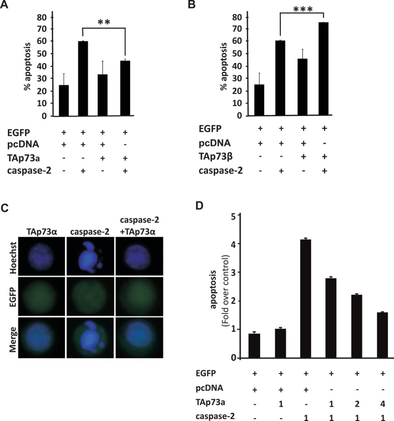
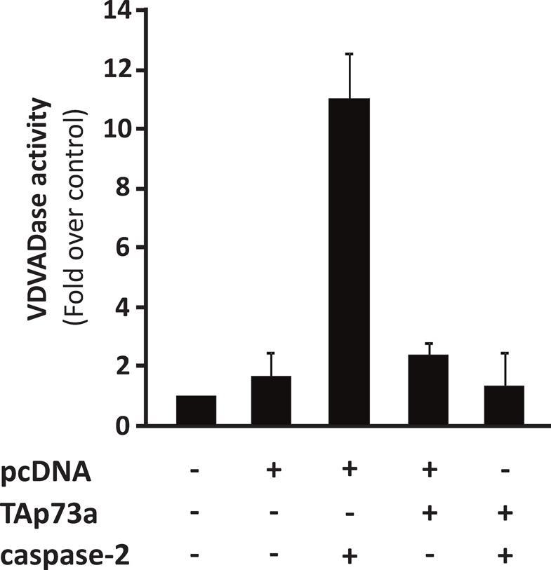
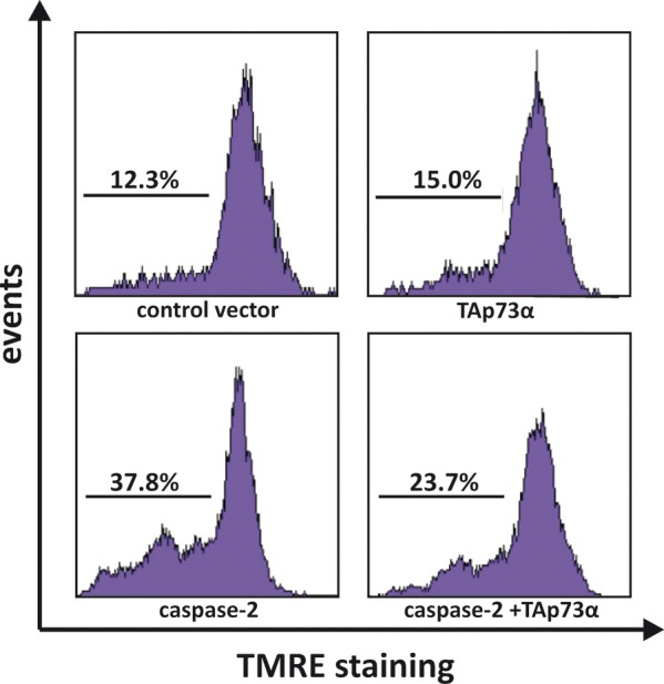
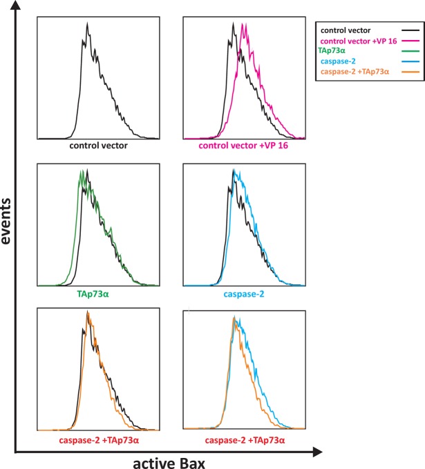
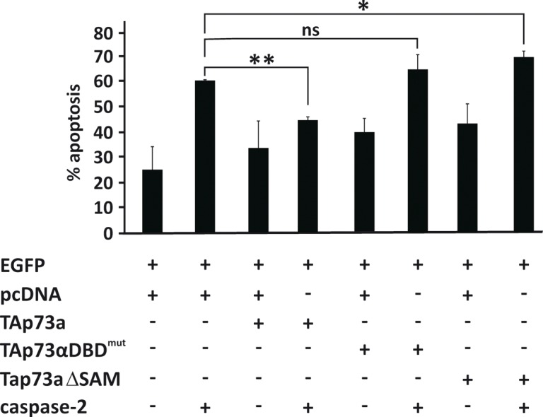
Caspase-2 is ubiquitously expressed and the most evolutionarily conserved mammalian caspase. It can be activated by a range of death stimuli prior to Bax activation and the occurrence of apoptotic mitochondrial dysfunctions. Caspase-2 has also been reported to exert tumour suppressor function in vivo. The full length TAp73alpha isoform is found up-regulated in various tumour types, and is reported in a cell-type specific manner to repress drug-induced apoptosis. Here, we report that TAp73alpha represses caspase-2 enzymatic activity and by this means reduce caspase-2 induced Bax activation, loss of mitochondrial transmembrane potential and resulting apoptosis. The inhibitory effect on caspase-2 requires the presence of the DNA binding domain and SAM domain region of TAp73alpha. In conclusion, the ability of TAp73alpha to act as an inhibitor of caspase-2-induced cell death together with its up-regulation in certain tumour types strengthen the potential oncogenic activities for this protein.


Conflict of interest statement

The authors confirm that there are no conflicts of interest.

Figures

Figure 1
Figure 1. TAp73alpha inhibits apoptosis induced by caspase-2 over-expression in SCLC NCI-H82 cells
SCLC NCI-H82 cells (Figure 1A-D) were co-transfected with EGFP together with either an empty (control) vector or plasmids encoding TAp73alpha, TAp73beta and/or caspase-2. Twenty-four hours post-transfection, cells were fixed in 4% PFA, stained with Hoechst and scored under a fluorescence microscope as percentage of EGFP expressing cells displaying condensed or fragmented nuclei. Three hundred EGFP transfected cells were analyzed and each experiment was repeated three times. Figures are mean + SD of three independent experiments, where ***p-value <0.001, **p-value <0.01
Figure 2
Figure 2. TAp73alpha expression inhibits caspase-2 activity
SCLC NCI-H82 cells (Figure 2) were transfected with TAp73alpha, caspase-2 and/or empty expression vectors, and caspase-2 activity was monitored. The enzyme activity of caspase-2 (VDVADase activity) was evaluated as the release of AMC from the substrate Ac-VDVAD-AMC (Ac-Val-Asp-Val-Ala-Asp-7-amino-4-methylcoumarin). The data represents three independent experiments.
Figure 3
Figure 3. TAp73alpha prevents caspase-2 induced mitochondrial dysfunction in SCLC NCI-H82 cells
SCLC NCI-H82 cells (Figure 3) were co-transfected with TAp73alpha, caspase-2 and/or empty expression vectors. Twenty-four hours post-transfection, cells were stained with TMRE and loss of mitochondrial trans-membrane potential (TMRE-negative cells) was assayed by scoring of EGFP transfected cells without TMRE by FACS analysis. Images are representatives of three independent experiments.
Figure 4
Figure 4. TAp73alpha counteracts caspase-2 induced Bax activation in SCLC NCI-H82 cells
SCLC NCI-H82 cells (Figure 4) were co-transfected with EGFP and plasmids encoding TAp73alpha, caspase-2 and/or empty expression vectors. Twenty-four hours post-transfection, SCLC NCI-H82 cells were collected, fixed in 4% PFA and incubated with the 6A7 anti-Bax antibody at 4°C, overnight, followed by secondary Alexa Fluor 635-conjugated antibody. For each sample 10,000 cells were sorted for green (EGFP) fluorescence using BD LSR II flow cytometer, and assessed for intensity of active Bax staining.
Figure 5
Figure 5. The DNA binding domain and SAM domain are required for TAp73alpha inhibition of caspase-2 induced apoptosis
SCLC NCI-H82 cells (Figure 5) were co-transfected with EGFP together with either an empty (control) vector, TAp73alpha or plasmids encoding different mutant forms of TAp73alpha (DBDmutp73alpha/p73alphaSAMdel), and/or caspase-2. Twenty-four hours post-transfection, cells were fixed, stained with Hoechst and scored under a fluorescence microscope as percentage of EGFP expressing cells displaying condensed or fragmented nuclei. Three hundred EGFP transfected cells were analyzed and each experiment was repeated three times. Figures are mean + SD of three independent experiments, where *p <0.05.

Similar articles

Cited by

References

    1. Tomasini R, Seux M, Nowak J, Bontemps C, Carrier A, Dagorn JC, Pébusque MJ, Iovanna JL, Dusetti NJ. TP53INP1 is a novel p73 target gene that induces cell cycle arrest and cell death by modulating p73 transcriptional activity. Oncogene. 2005;24:8093–8104. - PubMed
    1. Dobbelstein M, Strano S, Roth J, Blandino G. p73-induced apoptosis: a question of compartments and cooperation. Biochem Biophys Res Commun. 2005;331:688–693. - PubMed
    1. Marqués-García F, Ferrandiz N, Fernández-Alonso R, González-Cano L, Herreros-Villanueva M, Rosa-Garrido M, Fernández-García B, Vaque JP, Marqués MM, Alonso ME, Segovia JC, León J, Marín MC, et al. p73 plays a role in erythroid differentiation through GATA1 induction. J Biol Chem. 2009;284:21139–21156. - PMC - PubMed
    1. Meyer G, Cabrera Socorro A, Perez Garcia CG, Martinez Millan L, Walker N, Caput D. Developmental roles of p73 in Cajal-Retzius cells and cortical patterning. J Neurosci. 2004;24:9878–9887. - PMC - PubMed
    1. Yang A, Kaghad M, Caput D, McKeon F. On the shoulders of giants: p63, p73 and the rise of p53. Trends Genet. 2002;18:90–95. - PubMed

Publication types

MeSH terms