. 2012 Jan;122(1):70-9.
doi: 10.1172/JCI43713. Epub 2011 Dec 27.

Genetic inactivation of IL-1 signaling enhances atherosclerotic plaque instability and reduces outward vessel remodeling in advanced atherosclerosis in mice

Affiliations

Genetic inactivation of IL-1 signaling enhances atherosclerotic plaque instability and reduces outward vessel remodeling in advanced atherosclerosis in mice

Matthew R Alexander et al. J Clin Invest. 2012 Jan.

Erratum in

  • J Clin Invest. 2012 Feb 1;122(2):783

Abstract

Clinical complications of atherosclerosis arise primarily as a result of luminal obstruction due to atherosclerotic plaque growth, with inadequate outward vessel remodeling and plaque destabilization leading to rupture. IL-1 is a proinflammatory cytokine that promotes atherogenesis in animal models, but its role in plaque destabilization and outward vessel remodeling is unclear. The studies presented herein show that advanced atherosclerotic plaques in mice lacking both IL-1 receptor type I and apolipoprotein E (Il1r1⁻/⁻Apoe⁻/⁻ mice) unexpectedly exhibited multiple features of plaque instability as compared with those of Il1r1⁺/⁺Apoe⁻/⁻ mice. These features included reduced plaque SMC content and coverage, reduced plaque collagen content, and increased intraplaque hemorrhage. In addition, the brachiocephalic arteries of Il1r1⁻/⁻Apoe⁻/⁻ mice exhibited no difference in plaque size, but reduced vessel area and lumen size relative to controls, demonstrating a reduction in outward vessel remodeling. Interestingly, expression of MMP3 was dramatically reduced within the plaque and vessel wall of Il1r1⁻/⁻Apoe⁻/⁻ mice, and Mmp3⁻/⁻Apoe⁻/⁻ mice showed defective outward vessel remodeling compared with controls. In addition, MMP3 was required for IL-1-induced SMC invasion of Matrigel in vitro. Taken together, these results show that IL-1 signaling plays a surprising dual protective role in advanced atherosclerosis by promoting outward vessel remodeling and enhancing features of plaque stability, at least in part through MMP3-dependent mechanisms.


Figures

Figure 1
Figure 1. IL-1R1 deficiency reduces advanced atherosclerotic plaque size at the aortic root.
(A) Representative images of Movat-stained aortic sections of Il1r1–/–Apoe–/– and Il1r1+/+Apoe–/– mice after 27–30 weeks of high-fat diet feeding. Scale bars: 500 μm. (B) Quantification of total atherosclerotic plaque area within the aortic root of Il1r1+/+Apoe–/– and Il1r1–/–Apoe–/– mice at 150-μm intervals from the aortic valve attachment site. *P < 0.001 for difference between genotypes by Scheirer-Ray-Hare test. n = 13, Il1r1+/+Apoe–/–; n = 12, Il1r1–/–Apoe–/–. Data represent mean ± SEM.
Figure 2
Figure 2. IL-1R1 deficiency reduces compensatory outward remodeling of atherosclerotic brachiocephalic arteries.
(A) Movat staining of representative brachiocephalic arteries of Il1r1–/–Apoe–/– and Il1r1+/+Apoe–/– mice. Scale bars: 200 μm. (BD) Atherosclerotic plaque area (B), vessel area within the IEL (C), and lumen area, P < 0.001 for difference between genotypes by 2-way ANOVA, (D) at multiple locations along the brachiocephalic arteries of Il1r1–/–Apoe–/– and Il1r1+/+Apoe–/– mice, P < 0.001 for difference between genotypes by 2-way ANOVA after square root transformation. n = 14, Il1r1+/+Apoe–/–; n = 12, Il1r1–/–Apoe–/–. Data in BD represent mean ± SEM.
Figure 3
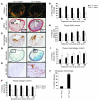
Figure 3. IL-1R1 deficiency decreases features of atherosclerotic plaque stability.
(AE) Representative images from brachiocephalic artery lesions of Il1r1+/+Apoe–/– and Il1r1–/–Apoe–/– mice with (A) picrosirius red staining and polarized light microscopy for collagen detection, (B) SM α-actin immunostaining for detecting SMCs on the plaque luminal surface (arrowheads) and total plaque SMC content, (C) Mac2 immunostaining for detection of plaque macrophages, (D) Movat staining for intraplaque rbc (arrow), and (E) immunostaining for the rbc marker TER-119 (magnified from boxed area in D). (FJ) Quantification of (F) plaque collagen content based on picrosirius red staining, P < 0.001 for difference of genotypes by 2-way ANOVA, (G) plaque SMC coverage based on SM a-actin staining, P < 0.001 for difference of genotypes by the Scheirer-Ray-Hare test, (H) total plaque SMC content based on SM a-actin staining, P < 0.001 for difference of genotypes by the Scheirer-Ray-Hare test (I) plaque macrophage content based on Mac2 staining, P = 0.01 for difference of genotypes by 2-way ANOVA after log transformation, and (J) the percentage of brachiocephalic arteries exhibiting intraplaque hemorrhage based on Movat and TER-119 staining, **P < 0.01 by Fisher’s exact test. Data in FI represent mean ± SEM. n = 14, Il1r1+/+Apoe–/–; n = 12, Il1r1–/–Apoe–/–. Scale bars: 200 μm (AD); 20 μm (inset, B; E).
Figure 4
Figure 4. IL-1R1 deficiency reduces MMP3 expression within the brachiocephalic artery plaque and vessel wall.
(A) Representative images of MMP3 staining within atherosclerotic brachiocephalic arteries just beyond the junction with the aortic arch from Il1r1+/+Apoe–/– and Il1r1–/–Apoe–/– mice. Scale bars: 200 μm. (B and C) Quantification of the percentage of positive area within the plaque (B) and vessel media (C) for MMP3 from Il1r1+/+Apoe–/– and Il1r1–/–Apoe–/– mice. *P < 0.001 by Mann-Whitney rank sum test. Data in B represent mean ± SEM (n = 14, Il1r1+/+Apoe–/–; n = 10, Il1r1–/–Apoe–/–), and data in C represent mean ± SEM (n = 14, Il1r1+/+Apoe–/–; n = 12, Il1r1–/–Apoe–/–).
Figure 5
Figure 5. IL-1β promotes MMP3-dependent SMC migration across a Matrigel barrier.
SMC number counted on the underside of Matrigel-coated Transwells was increased with IL-1β in the lower chamber in Mmp3+/+ SMCs, but not in Mmp3–/– SMCs. *P < 0.05 for IL-1β (50 ng/ml) versus vehicle by nested ANOVA. Data represent mean ± SEM of 3 independent experiments.

Comment in

References

    1. Lusis AJ. Atherosclerosis. Nature. 2000;407(6801):233–241. - PMC - PubMed
    1. Libby P, Theroux P. Pathophysiology of coronary artery disease. Circulation. 2005;111(25):3481–3488. doi: 10.1161/CIRCULATIONAHA.105.537878. - DOI - PubMed
    1. Galkina E, Ley K. Immune and inflammatory mechanisms of atherosclerosis. Annu Rev Immunol. 2009;27:165–197. doi: 10.1146/annurev.immunol.021908.132620. - DOI - PMC - PubMed
    1. Tedgui A, Mallat Z. Cytokines in atherosclerosis: pathogenic and regulatory pathways. Physiol Rev. 2006;86(2):515–581. doi: 10.1152/physrev.00024.2005. - DOI - PubMed
    1. Libby P, Ridker PM, Hansson GK. Inflammation in atherosclerosis: from pathophysiology to practice. . J Am Coll Cardiol. 2009;54(23):2129–2138. doi: 10.1016/j.jacc.2009.09.009. - DOI - PMC - PubMed

Publication types

MeSH terms