. 2012 Jan;122(1):267-79.
doi: 10.1172/JCI44927. Epub 2011 Dec 27.

Deletion of thioredoxin-interacting protein in mice impairs mitochondrial function but protects the myocardium from ischemia-reperfusion injury

Affiliations

Deletion of thioredoxin-interacting protein in mice impairs mitochondrial function but protects the myocardium from ischemia-reperfusion injury

Jun Yoshioka et al. J Clin Invest. 2012 Jan.

Abstract

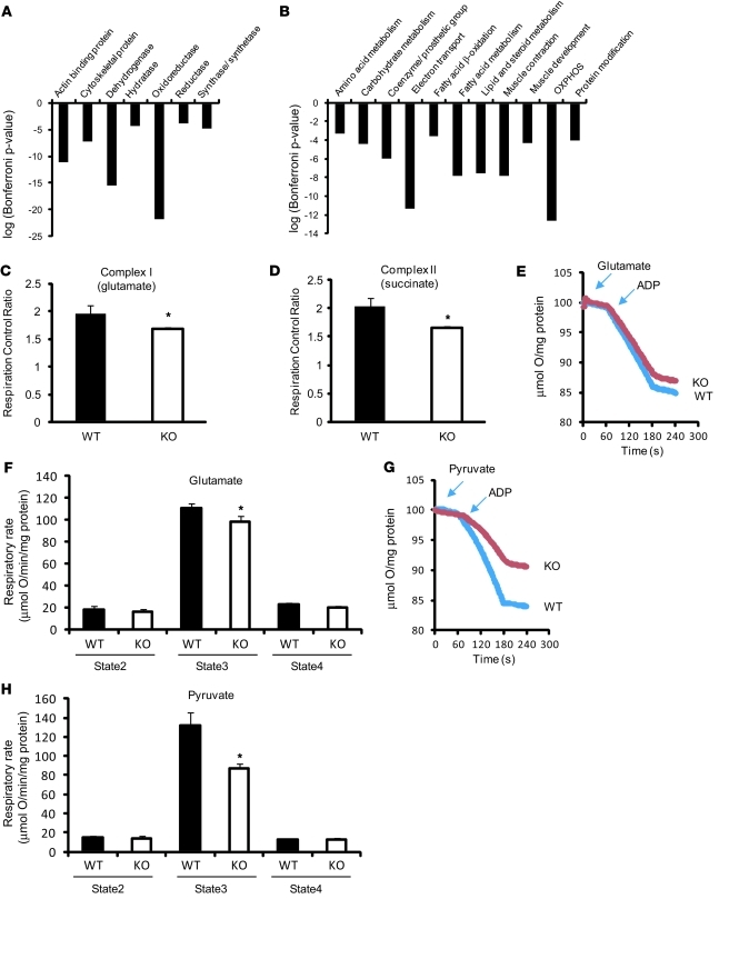
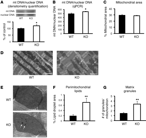
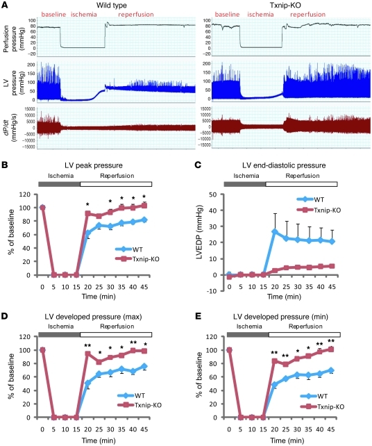
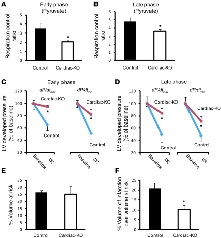
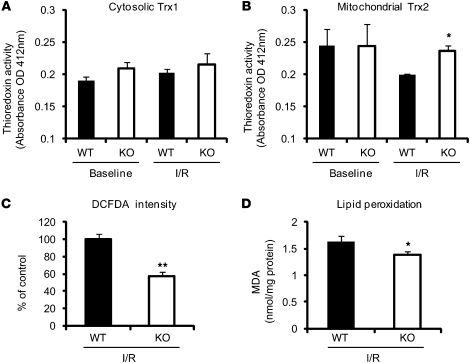
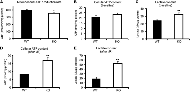
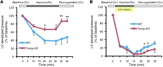
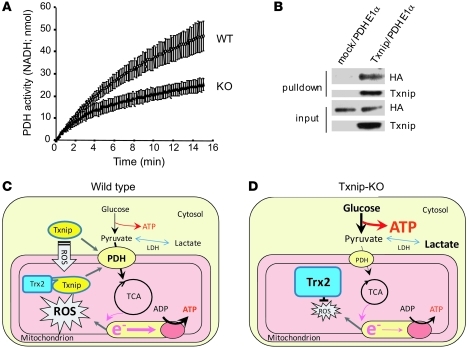
Classic therapeutics for ischemic heart disease are less effective in individuals with the metabolic syndrome. As the prevalence of the metabolic syndrome is increasing, better understanding of cardiac metabolism is needed to identify potential new targets for therapeutic intervention. Thioredoxin-interacting protein (Txnip) is a regulator of metabolism and an inhibitor of the antioxidant thioredoxins, but little is known about its roles in the myocardium. We examined hearts from Txnip-KO mice by polony multiplex analysis of gene expression and an independent proteomic approach; both methods indicated suppression of genes and proteins participating in mitochondrial metabolism. Consistently, Txnip-KO mitochondria were functionally and structurally altered, showing reduced oxygen consumption and ultrastructural derangements. Given the central role that mitochondria play during hypoxia, we hypothesized that Txnip deletion would enhance ischemia-reperfusion damage. Surprisingly, Txnip-KO hearts had greater recovery of cardiac function after an ischemia-reperfusion insult. Similarly, cardiomyocyte-specific Txnip deletion reduced infarct size after reversible coronary ligation. Coordinated with reduced mitochondrial function, deletion of Txnip enhanced anaerobic glycolysis. Whereas mitochondrial ATP synthesis was minimally decreased by Txnip deletion, cellular ATP content and lactate formation were higher in Txnip-KO hearts after ischemia-reperfusion injury. Pharmacologic inhibition of glycolytic metabolism completely abolished the protection afforded the heart by Txnip deficiency under hypoxic conditions. Thus, although Txnip deletion suppresses mitochondrial function, protection from myocardial ischemia is enhanced as a result of a coordinated shift to enhanced anaerobic metabolism, which provides an energy source outside of mitochondria.


Figures

Figure 1
Figure 1. Deletion of Txnip in the myocardium results in mitochondrial dysfunction with decreased expression of transcripts encoding mitochondrial metabolism.
(A and B) PMAGE demonstrated transcriptional changes in mitochondrial metabolism–related pathways in Txnip-KO hearts. Gene ontology analysis was performed using the software PANTHER that classified downregulated genes by their molecular functions (A) and biological processes (B) based on published evidence. Biological process (B) covers the biological systems to which a protein contributes. Each group refers to a group of proteins with common function or process. (C and D) Respiration control ratio was obtained from permeabilized cardiac fibers incubated with indicated substrates in 11 WT and 7 Txnip-KO mice. (EH) Representative tracing of oxygen consumption in isolated mitochondria (E and G). Mitochondria (200 μg) were incubated with 5 mM glutamate and 2 mM malate (E and F; n = 14 [WT] 13 [Txnip-KO]) or with 10 mM pyruvate and 2 mM malate (G and H; n = 4 each). State 2 and 3 respirations were measured by a Clark-type electrode in the presence of respiratory substrates before and after the addition of 1 mM ADP. State 4 respiration was measured on depletion of ADP. Values are mean ± SEM. *P < 0.05 versus WT.
Figure 2
Figure 2. Ultrastructural and quantitative analyses of mitochondria.
(A) Relative mtDNA copy number in the heart, calculated as the ratio of COX II (mitochondria) to COX IV (nuclear) genes as determined by Southern blotting. The ratio, quantified by densitometry, is shown as percent of WT control (n = 6 each). (B) The COX I gene of the mtDNA and the NDUFV1 nuclear DNA gene were amplified by qPCR. Amplification curves were used to determine the relative mtDNA/nuclear DNA ratio in each sample (n = 6). (CG) Electron micrographs from WT and Txnip-KO hearts showed morphological differences in mitochondria. Arrows denote lipid droplets (D) and mitochondrial matrix granules (E). Scale bars: 2 μm (D); 500 nm (E). Values are mean ± SEM. *P < 0.05, **P < 0.01 versus WT.
Figure 3
Figure 3. Txnip-KO hearts have improved functional recovery after ischemia-reperfusion injury.
(A) Representative tracing of LV pressure in a WT mouse heart (n = 3) and a Txnip-KO mouse heart (n = 4) with a Langendorff perfusion system. (BE) LV peak pressure (B), LV end-diastolic pressure (LVEDP; C), and maximal (D) and minimal (E) LV developed pressure were measured during 15 minutes ischemia and 30 minutes reperfusion. Values are mean ± SEM. *P < 0.05, **P < 0.01 versus WT.
Figure 4
Figure 4. Inducible cardiac-specific Txnip-KO mice have cardiometabolic phenotypes similar to those of systemic Txnip-KO mice.
(A and B) Respiratory parameters were obtained from permeabilized cardiac fibers incubated with 10 mM pyruvate and 5 mM malate in cardiomyocyte-specific Txnip-KO mice (Cardiac-KO). Early and late phases represent time points at 4 (n = 6) and 15 (n = 7) weeks, respectively, after deletion of Txnip by 4-hydroxy-tamoxifen. (C and D) Cardiac-specific Txnip-KO hearts had significantly improved functional recovery after ischemia-reperfusion injury. LV developed pressure (dP/dtmax and dP/dtmin) were measured with a Langendorff perfusion system at baseline and after ischemia-reperfusuion injury (I/R) — at 5 minutes reperfusion after 20 minutes ischemia. Values are mean ± SEM. n = 3–6 per group. (E and F) LV volume at risk (E; P = NS) and infarct volume over volume at risk (F; P < 0.05) in cardiac-specific Txnip-KO mice (n = 9) and their controls (n = 5), with transient (30 minutes) ischemia followed by reperfusion (24 hours) using a reversible ligation of the distal portion of left anterior descending artery. Values are mean ± SEM. *P < 0.05 versus control.
Figure 5
Figure 5. Effect of Txnip deletion on thioredoxin activities and oxidative stress in the myocardium after 15 minutes ischemia and 30 minutes reperfusion.
(A and B) Thioredoxin activity was measured using an insulin disulfide reduction assay in the cytosolic (A) and mitochondria-enriched (B) fractions. (C) Frozen sections were stained with DCFDA (4 μmol/l) to assess the tissue levels of ROS, and DCFDA intensities in the myocardium were calculated. (D) Tissue levels of MDA were measured in whole heart homogenates. Values are mean ± SEM. *P < 0.05, **P < 0.01 versus WT.
Figure 6
Figure 6. Deletion of Txnip enhances basal and ischemia-reperfusion anaerobic glycolysis.
(A) Mitochondrial ATP production rate was measured kinetically in isolated mitochondria after the addition of 1 mM ADP in the presence of the respiratory substrates 5 mM glutamate and 2 mM malate, using a luciferase/luciferin assay. (BE) Cellular ATP (B and D) and lactate (C and E) levels were measured either (B and C) at baseline from excised hearts in WT or Txnip-KO mice or (D and E) from isolated hearts in an ex vivo ischemia (15 minutes) and reperfusion (30 minutes) injury model. Values are mean ± SEM. n = 6–12 per group. *P < 0.05, **P < 0.01 versus WT.
Figure 7
Figure 7. Txnip-KO hearts increase anaerobic metabolism under hypoxia.
(A) Txnip-KO hearts had significantly improved mechanical function during hypoxia (95% N2, 5% CO2) and recovery (95% O2, 5% CO2). Hearts were perfused in a Langendorff heart model system in the presence of glucose (5.5 mM), mixed long-chain fatty acids (0.4 mM bound to 1% albumin), DL-β-hydroxybutyrate (0.38 mM), lactate (1 mM), and insulin (50 μU/ml). (B) To inhibit cellular glycolysis, hearts were perfused with oxamate (50 mM), a competitive inhibitor of LDH. n = 3 per group. Values are mean ± SEM. *P < 0.05, **P < 0.01 versus WT.
Figure 8
Figure 8. Txnip is involved in a key switch from aerobic to anaerobic metabolism.
(A) PDH activity was measured in isolated mitochondria as the rate of NADH production at 340 nm. (B) The interaction between Txnip and PDHE1α was assessed using a Txnip pulldown assay. HA-tagged PDHE1α was pulled down with Txnip, indicative of an interaction between these proteins. (C and D) Mechanism by which Txnip deletion may achieve cardioprotection in ischemia-reperfusion injury. (C) In WT hearts, upon stimulation with ROS, Txnip translocates into mitochondria to bind to and deactivate mitochondrial Trx2. (D) In Txnip-KO hearts, Trx2 is not inhibited by Txnip, thereby scavenging ROS efficiently. Txnip deficiency also reprograms glucose metabolism to more anaerobic ATP production in association with PDH, maintaining energy homeostasis in cardiomyocytes under ischemia-reperfusion injury.

Comment in

References

    1. Smith SC., Jr Multiple risk factors for cardiovascular disease and diabetes mellitus. Am J Med. 2007;120(3 suppl 1):S3–S11. - PubMed
    1. Kim JA, Wei Y, Sowers JR. Role of mitochondrial dysfunction in insulin resistance. Circ Res. 2008;102(4):401–414. doi: 10.1161/CIRCRESAHA.107.165472. - DOI - PMC - PubMed
    1. Chen Q, Camara AK, Stowe DF, Hoppel CL, Lesnefsky EJ. Modulation of electron transport protects cardiac mitochondria and decreases myocardial injury during ischemia and reperfusion. Am J Physiol Cell Physiol. 2007;292(1):C137–C147. doi: 10.1152/ajpcell.00270.2006. - DOI - PubMed
    1. Yellon DM, Hausenloy DJ. Myocardial reperfusion injury. N Engl J Med. 2007;357(11):1121–1135. doi: 10.1056/NEJMra071667. - DOI - PubMed
    1. Yoshioka J, Schreiter ER, Lee RT. Role of thioredoxin in cell growth through interactions with signaling molecules. Antioxid Redox Signal. 2006;8(11–12):2143–2151. - PubMed

Publication types

MeSH terms