

Modelling and dosimetry for alpha-particle therapy
- PMID: 22201712
- PMCID: PMC4332831
- DOI: 10.2174/1874471011104030261
Modelling and dosimetry for alpha-particle therapy
Abstract
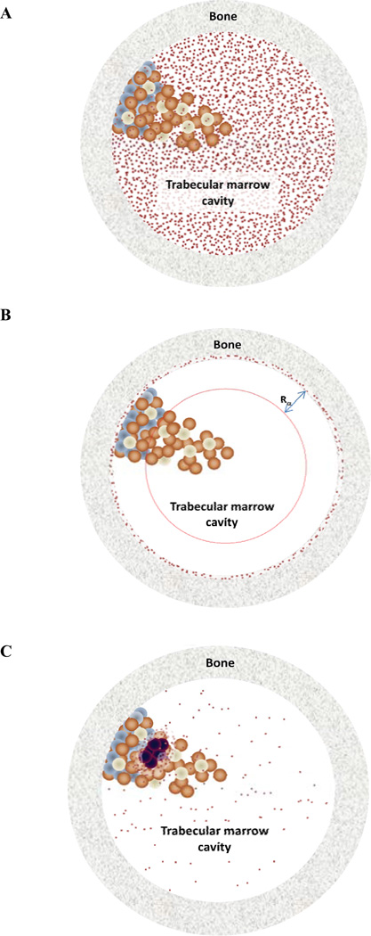
As a consequence of the high potency and short range of alpha-particles, radiopharmaceutical therapy with alpha- particle emitting radionuclides is a promising treatment approach that is under active pre-clinical and clinical investigation. To understand and predict the biological effects of alpha-particle radiopharmaceuticals, dosimetry is required at the micro or multi-cellular scale level. At such a scale, highly non-uniform irradiation of the target volume may be expected and the utility of a single absorbed dose value to predict biological effects comes into question. It is not currently possible to measure the pharmacokinetic input required for micro scale dosimetry in humans. Accordingly, pre-clinical studies are required to provide the pharmacokinetic data for dosimetry calculations. The translation of animal data to the human requires a pharmacokinetic model that links macro- and micro-scale pharmacokinetics thereby enabling the extrapolation of micro-scale kinetics from macroscopic measurements. These considerations along with a discussion of the appropriate physical quantity and related units for alpha-particle radiopharmaceutical therapy are examined in this review.
Figures
References
-
- Bolch WE, Eckerman KF, Sgouros G, Thomas SR. MIRD pamphlet No. 21: a generalized schema for radiopharmaceutical dosimetry--standardization of nomenclature. J. Nucl. Med. 2009;50(3):477–484. - PubMed
-
- Humm JL, Roeske JC, Fisher DR, Chen GT. Microdosimetric concepts in radioimmunotherapy. Med. Phys. 1993;20(2):535–541. - PubMed
-
- Roeske JC, Chen GT, Atcher RW, Pelizzari CA, Rotmensch J, Haraf D, Montag A, Weichselbaum RR. Modeling of dose to tumor and normal tissue from intraperitoneal radioimmunotherapy with alpha and beta emitters. Int. J. Radiat. Oncol. Biol. Phys. 1990;19(6):1539–1548. - PubMed
-
- Bäck T, Jacobsson L. The alpha-camera: a quantitative digital autoradiography technique using a charge-coupled device for ex vivo high-resolution bioimaging of alpha-particles. J. Nucl. Med. 2010;51(10):1616–1623. - PubMed
Publication types
MeSH terms
Substances
Grants and funding
LinkOut - more resources
Full Text Sources
Research Materials