Review
. 2012 Jan 1;17(1):375-88.
doi: 10.2741/3932.

Substrate channeling in proline metabolism

Affiliations
Review

Substrate channeling in proline metabolism

Benjamin W Arentson et al. Front Biosci (Landmark Ed). .

Abstract

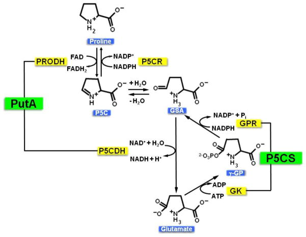
Proline metabolism is an important pathway that has relevance in several cellular functions such as redox balance, apoptosis, and cell survival. Results from different groups have indicated that substrate channeling of proline metabolic intermediates may be a critical mechanism. One intermediate is pyrroline-5-carboxylate (P5C), which upon hydrolysis opens to glutamic semialdehyde (GSA). Recent structural and kinetic evidence indicate substrate channeling of P5C/GSA occurs in the proline catabolic pathway between the proline dehydrogenase and P5C dehydrogenase active sites of bifunctional proline utilization A (PutA). Substrate channeling in PutA is proposed to facilitate the hydrolysis of P5C to GSA which is unfavorable at physiological pH. The second intermediate, gamma-glutamyl phosphate, is part of the proline biosynthetic pathway and is extremely labile. Substrate channeling of gamma-glutamyl phosphate is thought to be necessary to protect it from bulk solvent. Because of the unfavorable equilibrium of P5C/GSA and the reactivity of gamma-glutamyl phosphate, substrate channeling likely improves the efficiency of proline metabolism. Here, we outline general strategies for testing substrate channeling and review the evidence for channeling in proline metabolism.


Figures

Figure 1
Figure 1
Reactions of the proline metabolic pathway. In the catabolic pathway, proline is converted to glutamate via a four electron oxidation process. Proline dehydrogenase (PRODH) performs the first oxidative step, resulting in the intermediate pyrroline-5-carboxylate (P5C). P5C is subsequently hydrolyzed to glutamic semialdehyde (GSA), which is then further oxidized by P5C dehydrogenase (P5CDH) to generate glutamate. In Gram-negative bacteria, PRODH and P5CDH are fused together on a bifunctional enzyme called proline utilization A (PutA). Proline anabolism begins with phosphorylation of glutamate by gamma-glutamyl kinase (GK) to generate gamma-glutamyl phosphate (gamma-GP). gamma-GP is reduced by gamma-glutamyl phosphate reductase (GPR) to GSA, which cyclizes to form P5C. P5C is then reduced to proline via pyrroline-5-carboxylate reductase (P5CR). In higher eukaryotes such as plants and animals, GPR and GK are fused together in the bifunctional enzyme pyrroline-5-carboxylate synthase (P5CS).
Figure 2
Figure 2
Domain mapping of PRODH and P5CDH from E. coli (EcPutA), B. japonicum (BjPutA), and T. thermophilus. In PutAs, the PRODH and P5CDH domains are connected by a linker region (L). Trifunctional PutAs such as EcPutA also have a DNA binding domain (D). TtPRODH and TtP5CDH are separate enzymes (monofunctional) in the Gram-positive bacteria, T. thermophilus.
Figure 3
Figure 3
Domain mapping of monofunctional gamma-glutamyl phosphate reductase (EcGPR) and gamma-glutamyl kinase (EcGK) enzymes from E. coli and bifunctional pyrroline-5-carboxylate synthase (P5CS) from Homo sapiens. M, putative mitochondrial signaling peptide, BD, binding domain for glutamate and ATP, O, oligomerization domain, and PUA, pseudo uridine synthase and archaeosine-specific transglycosylase domain with no known function in EcGK.
Figure 4
Figure 4
Side reactions of intermediates pyrroline-5-carboxylate (P5C) and gamma-glutamyl phosphate (gamma-GP). (A) P5C can deactivate pyridoxal phosphate by forming an adduct, resulting in vitamin B6 deficiency in individuals with hyperprolinemia type II. The P5C-pyridoxal phosphate adduct structure is from reference . (B) gamma-GP can cyclize and dephosphorylate to form 5-oxoproline, which is suggested to be a neurotoxin in rats.
Figure 5
Figure 5
Strategies for examining substrate channeling. (A) Transient time analysis of a coupled reaction involving two enzymes, E1 and E2, which convert substrate A into product C. A trapping agent can also be used to test whether intermediate B is released into bulk solvent during the reaction. (B) Inactivation of one of the enzyme pairs by site-directed mutagenesis. If channeling occurs, adding inactive E2 would disrupt the active E1–E2 complex resulting in lower steady-state activity. (C) Testing channeling in bifunctional enzymes. Inactivation of the individual domains results in monofunctional variants that can only catalyze the coupled reaction via a diffusion mechanism. The mixture of monofunctional variants is thus a non-channeling control.
Figure 6
Figure 6
Structure of dimeric BjPutA shown in ribbon representation. The PRODH domain (red) and the P5CDH domain (orange) of each protomer are connected by a linker region (green). Active site residues (Arg456, Cys792), FAD and NAD+ are displayed as sticks. β-flap of each protomer is colored as magenta. The substrate channel of each BjPutA protomer is shown as blue surface. This model was made using PyMol, CAVER and PDB 3HAZ.
Figure 7
Figure 7
Example of transient time analysis of BjPutA. Steady-state formation of NADH using proline as a substrate by native BjPutA (solid black curve) and an equimolar mixture of monofunctional variants R456M and C792A (solid grey curve). The mixture of the monofunctional variants serves as a non-channeling control. The dotted line represents the extrapolation used for estimating the lag-time. Native BjPutA shows no apparent lag in NADH formation, while a lag time of about 6.5 min is observed for the non-channeling control. The dashed line overlaying the grey curve of the non-channeling control reaction was simulated using the kinetic parameters of PRODH and P5CDH as described previously and the following equation: [NADH] = v1t + (v1/v2)Km(ev2t/Km − 1) (21, 72). Assays were performed at pH 7.5.
Figure 8
Figure 8
Structures of GPR from T. maritima (TmGPR) and GK from E. coli (EcGK). EcGK is shown as a dimer with one monomer shown in surface representation and the other monomer as a ribbon cartoon illustration. Glutamate is shown as spheres in the substrate binding pocket, which is solvent accessible. Only one monomer of GPR (open conformation) is shown, which contains three domains: NADPH binding domain (yellow), catalytic domain (blue) with the catalytic cysteine shown in spheres, and the oligomerization domain (black). The solvent-exposed glutamate binding pocket of GK suggests that the gamma-glutamyl phosphate intermediate would be accessible to GPR in a potential GK-GPR complex. A GK-GPR complex in which the catalytic domain of GPR is aligned with the glutamate binding pocket of GK has been proposed and modeled by Marco-Marin et al. (32). Models shown here were made using PyMol and PDBs 2J5T (EcGK) and 1O20 (TmGPR).

Similar articles

Cited by

References

    1. Phang JM, Liu W, Zabirnyk O. Proline metabolism and microenvironmental stress. Annu Rev Nutr. 2010;30:441–463. - PMC - PubMed
    1. Donald SP, Sun XY, Hu CA, Yu J, Mei JM, Valle D, Phang JM. Proline oxidase, encoded by p53-induced gene-6, catalyzes the generation of proline-dependent reactive oxygen species. Cancer Res. 2001;61(5):1810–1815. - PubMed
    1. Krishnan N, Dickman MB, Becker DF. Proline modulates the intracellular redox environment and protects mammalian cells against oxidative stress. Free Radic Biol Med. 2008;44(4):671–681. - PMC - PubMed
    1. Chakravarti A. A compelling genetic hypothesis for a complex disease: PRODH2/DGCR6 variation leads to schizophrenia susceptibility. Proc Natl Acad Sci U S A. 2002;99(8):4755–4756. - PMC - PubMed
    1. Baumgartner MR, Hu CA, Almashanu S, Steel G, Obie C, Aral B, Rabier D, Kamoun P, Saudubray JM, Valle D. Hyperammonemia with reduced ornithine, citrulline, arginine and proline: a new inborn error caused by a mutation in the gene encoding delta(1)-pyrroline-5-carboxylate synthase. Hum Mol Genet. 2000;9(19):2853–2858. - PubMed

Publication types

MeSH terms

LinkOut - more resources