Review
. 2012 Jan 1;17(2):556-68.
doi: 10.2741/3943.

Unique structural features and sequence motifs of proline utilization A (PutA)

Affiliations
Review

Unique structural features and sequence motifs of proline utilization A (PutA)

Ranjan K Singh et al. Front Biosci (Landmark Ed). .

Abstract

Proline utilization A proteins (PutAs) are bifunctional enzymes that catalyze the oxidation of proline to glutamate using spatially separated proline dehydrogenase and pyrroline-5-carboxylate dehydrogenase active sites. Here we use the crystal structure of the minimalist PutA from Bradyrhizobium japonicum (BjPutA) along with sequence analysis to identify unique structural features of PutAs. This analysis shows that PutAs have secondary structural elements and domains not found in the related monofunctional enzymes. Some of these extra features are predicted to be important for substrate channeling in BjPutA. Multiple sequence alignment analysis shows that some PutAs have a 17-residue conserved motif in the C-terminal 20-30 residues of the polypeptide chain. The BjPutA structure shows that this motif helps seal the internal substrate-channeling cavity from the bulk medium. Finally, it is shown that some PutAs have a 100-200 residue domain of unknown function in the C-terminus that is not found in minimalist PutAs. Remote homology detection suggests that this domain is homologous to the oligomerization beta-hairpin and Rossmann fold domain of BjPutA.


Figures

Figure 1
Figure 1
The reactions of proline catabolism.
Figure 2
Figure 2
Phylogenetic tree representing the organization of proline catabolic enzymes in bacteria and eukaryotes. PutAs are found in branches 1 and 2. Monofunctional PRODH and P5CDH enzymes are found in branch 3. A cluster of trifunctional PutAs is indicated.
Figure 3
Figure 3
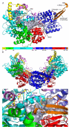
Structure of BjPutA. (A) Protomer structure with the domains colored according to the domain diagram. The silver surface represents the substrate-channeling cavity. FAD and NAD+ are represented as yellow and green sticks, respectively. Abbreviations used in the domain diagram are as follows: NBD, NAD+-binding domain; BH, beta-hairpin; CCM, conserved C-terminal motif. (B) The domain-swapped dimer of BjPutA. The domains are colored according to the domain diagram in panel A. The silver surfaces represent the substrate-channeling cavities of the two protomers. (C) Close-up view of the oligomerization domain covering the cavity of the other protomer. This figure and others were prepared with PyMol (72).
Figure 4
Figure 4
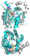
Comparison of the monofunctional enzyme TtPRODH (white) and the PRODH barrel of BjPutA (cyan). Strands of the barrel are labeled 1–8 using the standard convention for (beta-alpha)8 barrels. The extra helix of the PutA barrel is denoted alpha5a. Trp346 of alpha5a is colored magenta. The three helices of TtPRODH that precede the barrel are labeled alphaA, alphaB, and alphaC. Two orthogonal views are shown.
Figure 5
Figure 5
Comparison of the monofunctional enzyme TtP5CDH (white) and the P5CDH half of BjPutA. BjPutA is colored according to the legend in Figure 3A. NAD+ is drawn in green sticks.
Figure 6
Figure 6
Section of an MSA of branch 1 PutAs showing the conserved motif at the C-terminus. The trifunctional PutAs are EcPutA through Acidiphilium, plus Wigglesworthia. The other PutAs are bifunctional. The secondary structure elements are from the BjPutA structure (PDB code 3haz). This figure and others were prepared with ClustalW2 (73) and ESPript (74).
Figure 7
Figure 7
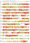
MSA of three branch 1 PutAs: BjPutA (Bj, GenBank BAC52526.1), Azoarcus sp. BH72 PutA (Az, GenBank CAL96369.1), and EcPutA (Ec, GenBank AAB59985.1). BjPutA is a minimalist PutA. Az is a long bifunctional PutA. Ec is a trifunctional PutA. The secondary structure elements above the sequence blocks are from the BjPutA structure (PDB code 3haz). The secondary structure elements for the N-terminal ribbon-helix-helix domain are from a structure of the EcPutA DNA-binding domain (PDB code 2gpe). Symbols below the sequence blocks denote the following: green squares, substrate-channeling cavity; triangles, proline binding site; hexagons, FAD binding site; diamonds, NAD+ binding site; ovals, GSA binding site; stars, catalytic Cys of the P5CDH catalytic domain and the Glu that is predicted to assist in hydrolysis of the thioacylenzyme.
Figure 8
Figure 8
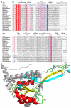
Homology of the CTD to the BjPutA beta-hairpin and Rossmann fold domain. (A) MSA of BjPutA with the CTDs of several branch 1 PutAs. (B) The structure of the NAD+-binding and oligomerization domains of BjPutA. The core region of homology with the CTD (residues 629–760) is colored according to secondary structure, with alpha helices in red, beta strands in yellow, and loops in green. Conserved residues of the CTD are indicated. Residues 510–628 are colored silver. Residues 955–989 are colored cyan.

Similar articles

Cited by

References

    1. Phang JM. The regulatory functions of proline and pyrroline-5-carboxylic acid. Curr. Top. Cell. Reg. 1985;25:92–132. - PubMed
    1. Ratzkin B, Roth J. Cluster of genes controlling proline degradation in Salmonella typhimurium. J. Bacteriol. 1978;133(2):744–54. - PMC - PubMed
    1. Tanner JJ. Structural biology of proline catabolism. Amino Acids. 2008;35(4):719–30. - PMC - PubMed
    1. Downing SJ, Phang JM, Kowaloff EM, Valle D, Smith RJ. Proline oxidase in cultured mammalian cells. J. Cell. Physiol. 1977;91(3):369–76. - PubMed
    1. Kowaloff EM, Phang JM, Granger AS, Downing SJ. Regulation of proline oxidase activity by lactate. Proc. Natl. Acad. Sci. U S A. 1977;74(12):5368–71. - PMC - PubMed

Publication types

MeSH terms

LinkOut - more resources