. 2012 Mar;15(1):33-45.
doi: 10.1007/s10456-011-9241-1. Epub 2011 Dec 22.

Exosomes released by K562 chronic myeloid leukemia cells promote angiogenesis in a Src-dependent fashion

Affiliations

Exosomes released by K562 chronic myeloid leukemia cells promote angiogenesis in a Src-dependent fashion

Marco Mineo et al. Angiogenesis. 2012 Mar.

Abstract

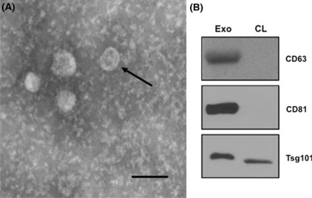
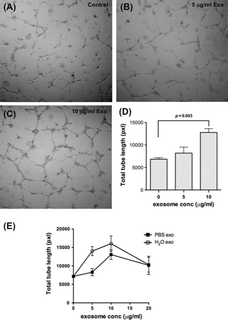
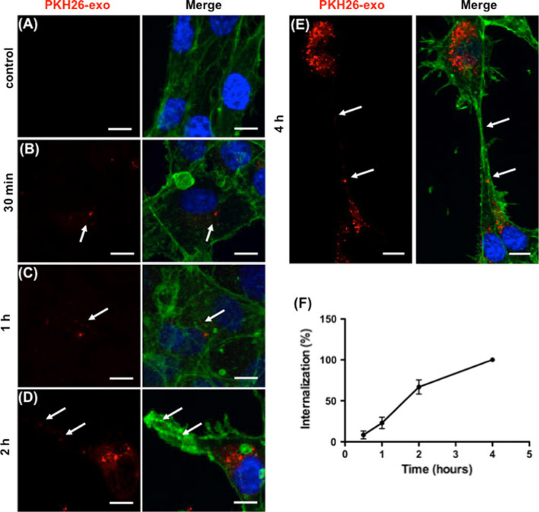
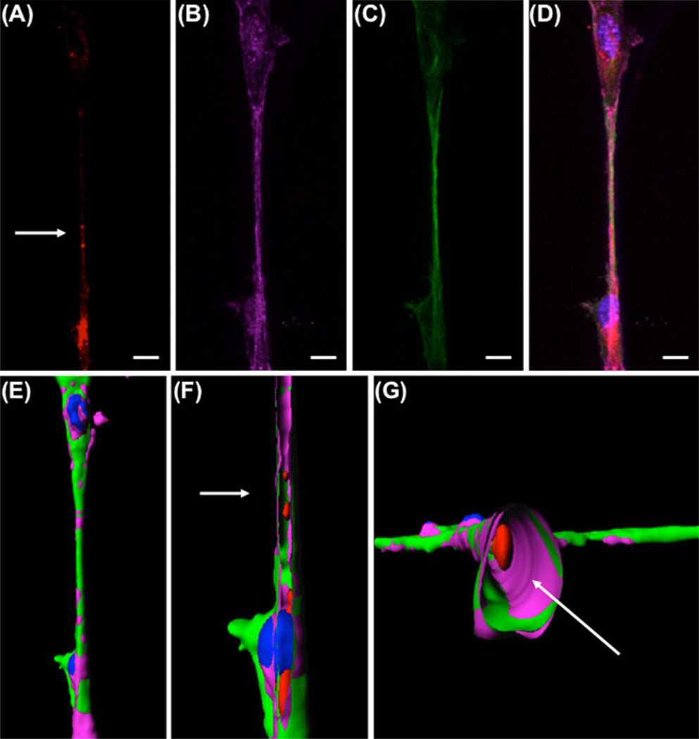
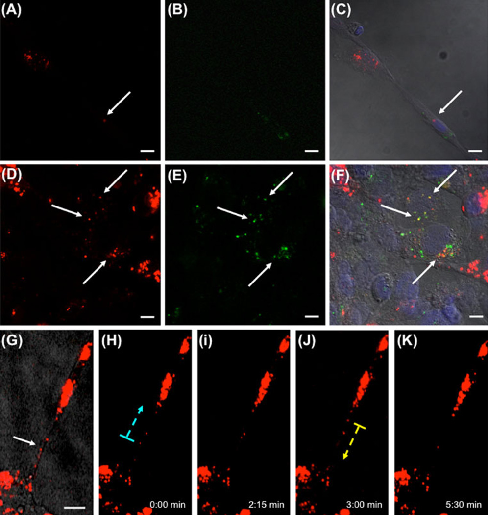
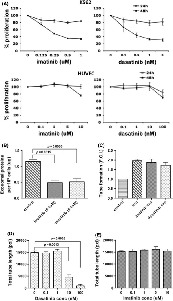
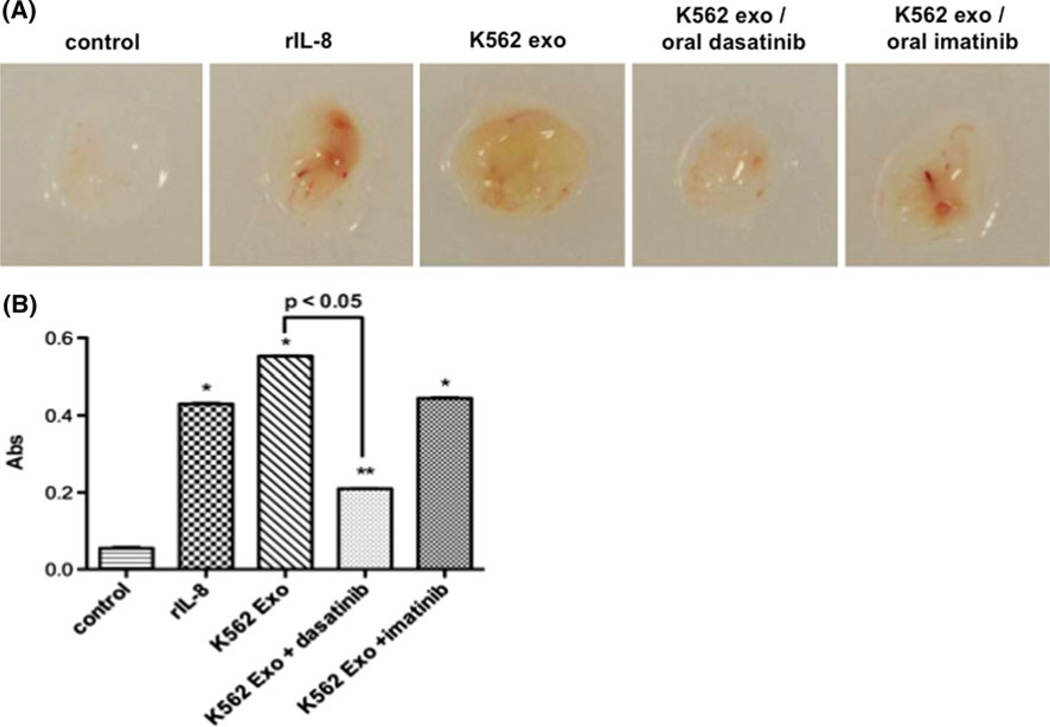
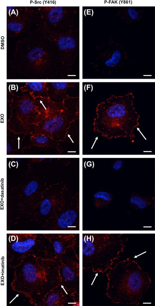
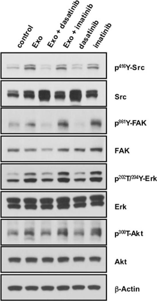
Exosomes, microvesicles of endocytic origin released by normal and tumor cells, play an important role in cell-to-cell communication. Angiogenesis has been shown to regulate progression of chronic myeloid leukemia (CML). The mechanism through which this happens has not been elucidated. We isolated and characterized exosomes from K562 CML cells and evaluated their effects on human umbilical endothelial cells (HUVECs). Fluorescent-labeled exosomes were internalized by HUVECs during tubular differentiation on Matrigel. Exosome localization was perinuclear early in differentiation, moving peripherally in cells undergoing elongation and connection. Exosomes move within and between nanotubular structures connecting the remodeling endothelial cells. They stimulated angiotube formation over a serum/growth factor-limited medium control, doubling total cumulative tube length (P = 0.003). Treatment of K562 cells with two clinically active tyrosine kinase inhibitors, imatinib and dasatinib, reduced their total exosome release (P < 0.009); equivalent concentrations of drug-treated exosomes induced a similar extent of tubular differentiation. However, dasatinib treatment of HUVECs markedly inhibited HUVEC response to drug control CML exosomes (P < 0.002). In an in vivo mouse Matrigel plug model angiogenesis was induced by K562 exosomes and abrogated by oral dasatinib treatment (P < 0.01). K562 exosomes induced dasatinib-sensitive Src phosphorylation and activation of downstream Src pathway proteins in HUVECs. Imatinib was minimally active against exosome stimulation of HUVEC cell differentiation and signaling. Thus, CML cell-derived exosomes induce angiogenic activity in HUVEC cells. The inhibitory effect of dasatinib on exosome production and vascular differentiation and signaling reveals a key role for Src in both the leukemia and its microenvironment.


Figures

Fig. 1
Fig. 1
K562 CML cells release exosomes in their CM. a Morphology of exosomes isolated from medium conditioned by K562 cells were analyzed by transmission electron microscopy. Arrow cup-shaped exosome. Scale bar 150 nm. b Exosome characterization by immunoblot. Equal amounts of total exosomal proteins (Exo) and K562 cell lysate (CL) were analyzed for the expression of CD63, CD81, and Tsg101
Fig. 2
Fig. 2
Dose dependent stimulation of vascular tube formation by K562 exosomes. a–c Exosomes promote tube formation by HUVECs on Matrigel. a control; b 5 µg/ml exosomes; c 10 µg/ml exosome. d Quantitative analysis of the total tube length. Data are mean ± SEM of triplicate experiments. e Lysed K562 exosomes are equipotent or more active than intact exosomes. Data are mean ± SEM of triplicate experiments
Fig. 3
Fig. 3
Time dependent uptake and localization of exosomes in HUVEC cells during differentiation. a–e Exosomes labeled with PKH26 (red) were added to HUVEC cells at time of plating and incubated as indicated. Effluent from a filtered suspension of PKH26- labeled exosomes was incubated with the HUVECs for 4 h as the control. Cells were fixed and stained for actin (green), and nuclei (DAPI, blue). Arrows indicate exosome localization. Scale bars 10 µm. f Quantitation of HUVEC uptake of PKH26-labeled K562 exosomes. The data represent the mean ± SEM of three independent experiments
Fig. 4
Fig. 4
Exosomes localize inside nanotubes. a–d HUVEC cells incubated with PKH26-labeled exosomes (a, red) on Matrigel reorganized and sent out pseudopods that extended into nanotubes containing both F-actin (b, pink) and microtubules (c, green). Nuclei were stained with DAPI (blue). The merged image (d) shows the localization of exosomes at level of the nanotube. Scale bars 10 µm. e 3D reconstruction of the nanotube by surface rendering, showing outer view. f–g Sections of the nanotube showing exosome localization inside the nanotubes in longitudinal section (f) and cross section (g)
Fig. 5
Fig. 5
Exosomes are transferred cell-to-cell. a–f HUVECs carrying PKH26-stained exosomes (a/d, red) were mixed with HUVECs carrying PKH67-stained exosomes (b/e green) and plated on Matrigel for 4 h. After the incubation, cells were fixed and DAPI stained for nuclei (blue). Tube forming cells show the presence of both green and red exosomes inside the same cell (c/f, arrow). The arrows in figures d–f point to red exosomes co-localizing with green exosomes. g–k Exosomes move along nanotubes. HUVECs were incubated with PKH26-labelled exosomes and analyzed by fluorescent time-lapse video microscopy. g Brightfield view of HUVECs connected by nanotubes carrying exosomes (arrows). h–k Selected frames of a video sequence showing transfer of PKH26-labeled exosomes (dotted arrows indicate direction of movement). Scale bars: 10 µm
Fig. 6
Fig. 6
CML therapy regulates exosome release and exosomeinduced tube formation. a Effects of imatinib and dasatinib on K562 and HUVEC proliferation. Imatinib and dasatinib exposure continued for 24 or 48 h. b Both imatinib and dasatinib reduce total released exosomal protein. c Exosomes from imatinib- or dasatinib-treated K562 cells (0.1 µM, and 0.1 nM, respectively) are equipotent as those from vehicle-treated cells. d, e A dose-dependent inhibition by dasatinib (d), but not imatinib (e) on the HUVEC response to control exosomes. Data are mean ± SEM of triplicate experiments
Fig. 7
Fig. 7
K562 exosomes induce vascularization of Matrigel plugs in vivo. a Nude mice were injected with Matrigel plugs containing PBS or recombinant IL-8 (positive control), or 100 µg/plug K562 exosomes. Mice were treated with oral administration of dasatinib or imatinib as described in Materials and Methods, and analyzed for plug vascularization after 14 days of treatment. b Plug hemoglobin concentration as a measure of vascularity. Comparison of IL-8 or K562 exosome-containing plugs vs control: * P ≤ 0.01, ** P <0.05
Fig. 8
Fig. 8
Dasatinib-sensitive exosome activation of Src signaling during HUVEC differentiation. HUVECs with 10 µg/ml control exosomes were plated on Matrigel in presence of DMSO vehicle control (a, e), dasatinib (10 nM), or imatinib (1 µM), and allowed to adhere for 30 min. Cells were fixed and red fluorescence labeling was performed to visualize p-Src (a–d) or p-FAK (e–h). Nuclei were stained with DAPI (blue). Scale bars 10 µm
Fig. 9
Fig. 9
Src-dependent exosome activation of signaling pathways in HUVECs. HUVECs growing in monolayer were incubated with or without exosomes and treated with DMSO (control), dasatnib (10 nM), or imatinib (1 µM) for 2 h. Data are representative of 2 or more blots

References

    1. Folkman J. Role of angiogenesis in tumor growth and metastasis. Semin Oncol. 2002;29:15–18. - PubMed
    1. Aguayo A, Kantarjian H, Manshouri T, Gidel C, Estey E, Thomas D, Koller C, Estrov Z, O’Brien S, Keating M, Freireich E, Albitar M. Angiogenesis in acute and chronic leukemias and myelodysplastic syndromes. Blood. 2000;96:2240–2245. - PubMed
    1. Lundberg LG, Lerner R, Sundelin P, Rogers R, Folkman J, Palmblad J. Bone marrow in polycythemia vera, chronic myelocytic leukemia, and myelofibrosis has an increased vascularity. Am J Pathol. 2000;157:15–19. - PMC - PubMed
    1. Zhelyazkova AG, Tonchev AB, Kolova P, Ivanova L, Gercheva L. Prognostic significance of hepatocyte growth factor and microvessel bone marrow density in patients with chronic myeloid leukaemia. Scand J Clin Lab Invest. 2008;68:492–500. - PubMed
    1. Nowell PC, Hungerford DA. A minute chromosome in human chronic granulocytic leukemia. Science. 1960;132:1497–1499.

Publication types

MeSH terms